 Movie
Movie Controller

[English] 日本語
 Yorodumi
- PDB-1mqs: Crystal structure of Sly1p in complex with an N-terminal peptide ... -
+ Open data

- Basic information
| Entry | Database: PDB / ID: 1mqs | ||||||
|---|---|---|---|---|---|---|---|
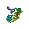
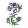
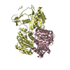
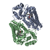
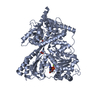
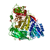
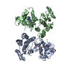
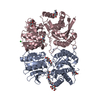
| Title | Crystal structure of Sly1p in complex with an N-terminal peptide of Sed5p | ||||||
 Components Components |
| ||||||
 Keywords Keywords | ENDOCYTOSIS/EXOCYTOSIS / SM-protein / SNARE / syntaxin / ENDOCYTOSIS-EXOCYTOSIS COMPLEX | ||||||
| Function / homology |  Function and homology information Function and homology informationregulation of inclusion body assembly / Cargo concentration in the ER / RHOC GTPase cycle / Golgi cis cisterna membrane / RHOA GTPase cycle / Insertion of tail-anchored proteins into the endoplasmic reticulum membrane / Intra-Golgi traffic / vesicle fusion with Golgi apparatus / regulation of Ras protein signal transduction / COPI-mediated anterograde transport ...regulation of inclusion body assembly / Cargo concentration in the ER / RHOC GTPase cycle / Golgi cis cisterna membrane / RHOA GTPase cycle / Insertion of tail-anchored proteins into the endoplasmic reticulum membrane / Intra-Golgi traffic / vesicle fusion with Golgi apparatus / regulation of Ras protein signal transduction / COPI-mediated anterograde transport / COPII-mediated vesicle transport / : / SNARE complex / SNAP receptor activity / vesicle fusion / cis-Golgi network / intra-Golgi vesicle-mediated transport / retrograde vesicle-mediated transport, Golgi to endoplasmic reticulum / COPII-coated ER to Golgi transport vesicle / syntaxin binding / positive regulation of SNARE complex assembly / endoplasmic reticulum to Golgi vesicle-mediated transport / endomembrane system / SNARE binding / intracellular protein transport / Golgi membrane / endoplasmic reticulum membrane / endoplasmic reticulum / membrane / cytoplasm / cytosol Similarity search - Function | ||||||
| Biological species |  | ||||||
| Method |  X-RAY DIFFRACTION / SYNCHROTRON / SAD / Resolution: 3 Å X-RAY DIFFRACTION / SYNCHROTRON / SAD / Resolution: 3 Å | ||||||
 Authors Authors | Bracher, A. / Weissenhorn, W. | ||||||
 Citation Citation | Journal: Embo J. / Year: 2002 Title: Structural basis for the Golgi membrane recruitment of Sly1p by Sed5p Authors: Bracher, A. / Weissenhorn, W. #1: Journal: Nature / Year: 2000Title: Three-dimensional structure of the neuronal-Sec1-syntaxin 1a complex Authors: Misura, K.M.S. / Scheller, R.H. / Weis, W.I. #2: Journal: Structure / Year: 2000Title: The X-ray crystal structure of neuronal Sec1 from squid sheds new light on the role of this protein in exocytosis Authors: Bracher, A. / Perrakis, A. / Dresbach, T. / Betz, H. / Weissenhorn, W. #3: Journal: J.Mol.Biol. / Year: 2001Title: Crystal structures of neuronal squid Sec1 implicate inter-domain hinge movement in the release of t-SNAREs Authors: Bracher, A. / Weissenhorn, W. | ||||||
| History |
|
- Structure visualization
| Structure viewer | Molecule: MolmilJmol/JSmol |
|---|
- Downloads & links
-Download
| PDBx/mmCIF format | 1mqs.cif.gz | 134.4 KB | Display | PDBx/mmCIF format |
|---|---|---|---|---|
| PDB format | pdb1mqs.ent.gz | 102.8 KB | Display | PDB format |
| PDBx/mmJSON format | 1mqs.json.gz | Tree view | PDBx/mmJSON format | |
| Others |  Other downloads Other downloads |
-Validation report
| Arichive directory | https://data.pdbj.org/pub/pdb/validation_reports/mq/1mqsftp://data.pdbj.org/pub/pdb/validation_reports/mq/1mqs | HTTPS FTP |
|---|
-Related structure data
| Related structure data | |
|---|---|
| Similar structure data |


-Links
 PDBj
PDBj


- Assembly
| Deposited unit | 
| ||||||||
|---|---|---|---|---|---|---|---|---|---|
| 1 |
| ||||||||
| 2 | 
| ||||||||
| Unit cell |
|
-Components
| #1: Protein | Mass: 75655.688 Da / Num. of mol.: 1 Source method: isolated from a genetically manipulated source Source: (gene. exp.) Plasmid: PMAL-TEV / Production host:  |
|---|---|
| #2: Protein/peptide | Mass: 5949.565 Da / Num. of mol.: 1 Source method: isolated from a genetically manipulated source Source: (gene. exp.) Plasmid: pETM30 / Production host: |
| #3: Water | ChemComp-HOH /  Mass: 18.015 Da / Num. of mol.: 20 / Source method: isolated from a natural source / Formula: H2O Mass: 18.015 Da / Num. of mol.: 20 / Source method: isolated from a natural source / Formula: H2O |
| Has protein modification | Y |
-Experimental details
-Experiment
| Experiment | Method: X-RAY DIFFRACTION / Number of used crystals: 1 |
|---|
- Sample preparation
| Crystal | Density Matthews: 3.5 Å3/Da / Density % sol: 64.89 % | |||||||||||||||||||||||||||||||||||
|---|---|---|---|---|---|---|---|---|---|---|---|---|---|---|---|---|---|---|---|---|---|---|---|---|---|---|---|---|---|---|---|---|---|---|---|---|
| Crystal grow | Temperature: 293 K / Method: vapor diffusion, hanging drop / pH: 7.2 Details: 3.5M sodium formate, 20mM Tris HCl pH 7.2, 50mM NaCl, VAPOR DIFFUSION, HANGING DROP, temperature 293K | |||||||||||||||||||||||||||||||||||
| Crystal grow | *PLUS | |||||||||||||||||||||||||||||||||||
| Components of the solutions | *PLUS
|
-Data collection
| Diffraction |
| |||||||||||||||
|---|---|---|---|---|---|---|---|---|---|---|---|---|---|---|---|---|
| Diffraction source | Source: SYNCHROTRON / Site: ESRF  / Beamline: BM14 / Wavelength: 0.9875,0.9793,0.9789,0.9184 / Beamline: BM14 / Wavelength: 0.9875,0.9793,0.9789,0.9184 | |||||||||||||||
| Detector |
| |||||||||||||||
| Radiation | Protocol: SINGLE WAVELENGTH / Monochromatic (M) / Laue (L): M / Scattering type: x-ray | |||||||||||||||
| Radiation wavelength |
| |||||||||||||||
| Reflection | Resolution: 2.9→30 Å / Num. all: 26182 / Num. obs: 26182 / % possible obs: 100 % / Observed criterion σ(F): 0 / Observed criterion σ(I): 0 / Redundancy: 7.7 % / Biso Wilson estimate: 92.3 Å2 / Rmerge(I) obs: 0.044 / Net I/σ(I): 13.3 | |||||||||||||||
| Reflection shell | Resolution: 2.9→3 Å / Rmerge(I) obs: 0.552 / % possible all: 100 | |||||||||||||||
| Reflection | *PLUS Lowest resolution: 30 Å / % possible obs: 100 % / Num. measured all: 200527 | |||||||||||||||
| Reflection shell | *PLUS % possible obs: 100 % |
- Processing
| Software |
| |||||||||||||||||||||||||
|---|---|---|---|---|---|---|---|---|---|---|---|---|---|---|---|---|---|---|---|---|---|---|---|---|---|---|
| Refinement | Method to determine structure: SAD / Resolution: 3→30 Å / Rfactor Rfree error: 0.008 / Isotropic thermal model: RESTRAINED / Cross valid method: THROUGHOUT / σ(F): 0 / σ(I): 0 / Stereochemistry target values: Engh & Huber
| |||||||||||||||||||||||||
| Solvent computation | Bsol: 52.0133 Å2 / ksol: 0.31282 e/Å3 | |||||||||||||||||||||||||
| Displacement parameters | Biso mean: 97.7 Å2
| |||||||||||||||||||||||||
| Refine analyze |
| |||||||||||||||||||||||||
| Refinement step | Cycle: LAST / Resolution: 3→30 Å
| |||||||||||||||||||||||||
| Refine LS restraints |
| |||||||||||||||||||||||||
| Xplor file |
| |||||||||||||||||||||||||
| Refinement | *PLUS Highest resolution: 3 Å / Lowest resolution: 30 Å | |||||||||||||||||||||||||
| Solvent computation | *PLUS | |||||||||||||||||||||||||
| Displacement parameters | *PLUS | |||||||||||||||||||||||||
| Refine LS restraints | *PLUS
|
