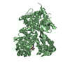
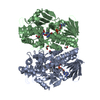
SHEET DETERMINATION METHOD: DSSP THE SHEETS PRESENTED AS "AD" IN EACH CHAIN ON SHEET RECORDS BELOW ... SHEET DETERMINATION METHOD: DSSP THE SHEETS PRESENTED AS "AD" IN EACH CHAIN ON SHEET RECORDS BELOW IS ACTUALLY AN 5-STRANDED BARREL THIS IS REPRESENTED BY A 6-STRANDED SHEET IN WHICH THE FIRST AND LAST STRANDS ARE IDENTICAL. THE SHEETS PRESENTED AS "BD" IN EACH CHAIN ON SHEET RECORDS BELOW IS ACTUALLY AN 5-STRANDED BARREL THIS IS REPRESENTED BY A 6-STRANDED SHEET IN WHICH THE FIRST AND LAST STRANDS ARE IDENTICAL.
Protocol: SINGLE WAVELENGTH / Monochromatic (M) / Laue (L): M / Scattering type: x-ray
Radiation wavelength
Wavelength: 0.9793 Å / Relative weight: 1
Reflection
Resolution: 1.9→102 Å / Num. obs: 160290 / % possible obs: 99.8 % / Observed criterion σ(I): 2 / Redundancy: 5.3 % / Biso Wilson estimate: 27.07 Å2 / Rmerge(I) obs: 0.09 / Net I/σ(I): 13.4
Reflection shell
Resolution: 1.9→2 Å / Redundancy: 4.1 % / Rmerge(I) obs: 0.6 / Mean I/σ(I) obs: 2.2 / % possible all: 98.6
-
Processing
Software
Name
Version
Classification
PHENIX
(PHENIX.REFINE)
refinement
MOSFLM
datareduction
SCALA
datascaling
autoSHARP
phasing
Refinement
Method to determine structure: SAD Starting model: NONE Resolution: 1.9→34.252 Å / SU ML: 0.26 / σ(F): 1.34 / Phase error: 21.62 / Stereochemistry target values: ML Details: ELECTRON DENSITY WAS NOT VISIBLE FOR RESIDUES 1-2 AND 750-767 AND 6-HISTIDINE TAG, PROBABLY DUE TO STRUCTURAL DISORDER, AND ARE THEREFORE ABSENT FROM THE MODEL
Rfactor
Num. reflection
% reflection
Rfree
0.224
8044
5 %
Rwork
0.1837
-
-
obs
0.1857
160181
99.72 %
Solvent computation
Shrinkage radii: 0.9 Å / VDW probe radii: 1.11 Å / Solvent model: FLAT BULK SOLVENT MODEL / Bsol: 40.242 Å2 / ksol: 0.4 e/Å3
Displacement parameters
Biso mean: 32.1 Å2
Baniso -1
Baniso -2
Baniso -3
1-
-2.5314 Å2
-0 Å2
0 Å2
2-
-
-2.5314 Å2
-0 Å2
3-
-
-
5.0627 Å2
Refinement step
Cycle: LAST / Resolution: 1.9→34.252 Å
Protein
Nucleic acid
Ligand
Solvent
Total
Num. atoms
11951
0
78
1419
13448
Refine LS restraints
Refine-ID
Type
Dev ideal
Number
X-RAY DIFFRACTION
f_bond_d
0.017
12279
X-RAY DIFFRACTION
f_angle_d
1.557
16628
X-RAY DIFFRACTION
f_dihedral_angle_d
18.298
4634
X-RAY DIFFRACTION
f_chiral_restr
0.104
1833
X-RAY DIFFRACTION
f_plane_restr
0.008
2170
LS refinement shell
Resolution (Å)
Rfactor Rfree
Num. reflection Rfree
Rfactor Rwork
Num. reflection Rwork
Refine-ID
% reflection obs (%)
1.9-1.9216
0.3164
250
0.2708
4778
X-RAY DIFFRACTION
95
1.9216-1.9442
0.2713
269
0.2604
4935
X-RAY DIFFRACTION
99
1.9442-1.9679
0.2643
234
0.2428
5034
X-RAY DIFFRACTION
100
1.9679-1.9928
0.2909
266
0.2343
5081
X-RAY DIFFRACTION
100
1.9928-2.019
0.2744
297
0.2105
4975
X-RAY DIFFRACTION
100
2.019-2.0467
0.2611
280
0.2171
4982
X-RAY DIFFRACTION
100
2.0467-2.0759
0.2568
243
0.2161
5081
X-RAY DIFFRACTION
100
2.0759-2.1069
0.2643
284
0.2031
5010
X-RAY DIFFRACTION
100
2.1069-2.1398
0.2333
296
0.2002
5032
X-RAY DIFFRACTION
100
2.1398-2.1749
0.2318
272
0.1891
5084
X-RAY DIFFRACTION
100
2.1749-2.2124
0.2715
247
0.1879
4984
X-RAY DIFFRACTION
100
2.2124-2.2526
0.226
271
0.1798
5068
X-RAY DIFFRACTION
100
2.2526-2.2959
0.2343
248
0.1813
5073
X-RAY DIFFRACTION
100
2.2959-2.3428
0.2169
263
0.1747
5070
X-RAY DIFFRACTION
100
2.3428-2.3937
0.243
262
0.1774
5065
X-RAY DIFFRACTION
100
2.3937-2.4494
0.2519
256
0.1784
5076
X-RAY DIFFRACTION
100
2.4494-2.5106
0.222
250
0.167
5066
X-RAY DIFFRACTION
100
2.5106-2.5785
0.2246
299
0.1731
5009
X-RAY DIFFRACTION
100
2.5785-2.6543
0.2283
256
0.1781
5087
X-RAY DIFFRACTION
100
2.6543-2.74
0.2657
258
0.1801
5094
X-RAY DIFFRACTION
100
2.74-2.8379
0.2424
255
0.1739
5087
X-RAY DIFFRACTION
100
2.8379-2.9514
0.2408
265
0.1811
5114
X-RAY DIFFRACTION
100
2.9514-3.0857
0.2329
270
0.1767
5094
X-RAY DIFFRACTION
100
3.0857-3.2482
0.215
285
0.1779
5078
X-RAY DIFFRACTION
100
3.2482-3.4516
0.2099
275
0.175
5122
X-RAY DIFFRACTION
100
3.4516-3.7178
0.1814
287
0.16
5099
X-RAY DIFFRACTION
100
3.7178-4.0914
0.186
268
0.1523
5145
X-RAY DIFFRACTION
100
4.0914-4.6821
0.1683
274
0.1426
5194
X-RAY DIFFRACTION
100
4.6821-5.8941
0.2086
284
0.1643
5209
X-RAY DIFFRACTION
100
5.8941-34.2572
0.2126
280
0.2059
5411
X-RAY DIFFRACTION
99
+
About Yorodumi
-
News
-
Feb 9, 2022. New format data for meta-information of EMDB entries
New format data for meta-information of EMDB entries
Version 3 of the EMDB header file is now the official format.
The previous official version 1.9 will be removed from the archive.
In the structure databanks used in Yorodumi, some data are registered as the other names, "COVID-19 virus" and "2019-nCoV". Here are the details of the virus and the list of structure data.
Jan 31, 2019. EMDB accession codes are about to change! (news from PDBe EMDB page)
EMDB accession codes are about to change! (news from PDBe EMDB page)
The allocation of 4 digits for EMDB accession codes will soon come to an end. Whilst these codes will remain in use, new EMDB accession codes will include an additional digit and will expand incrementally as the available range of codes is exhausted. The current 4-digit format prefixed with “EMD-” (i.e. EMD-XXXX) will advance to a 5-digit format (i.e. EMD-XXXXX), and so on. It is currently estimated that the 4-digit codes will be depleted around Spring 2019, at which point the 5-digit format will come into force.
The EM Navigator/Yorodumi systems omit the EMD- prefix.
Related info.:Q: What is EMD? / ID/Accession-code notation in Yorodumi/EM Navigator
Yorodumi is a browser for structure data from EMDB, PDB, SASBDB, etc.
This page is also the successor to EM Navigator detail page, and also detail information page/front-end page for Omokage search.
The word "yorodu" (or yorozu) is an old Japanese word meaning "ten thousand". "mi" (miru) is to see.
Related info.:EMDB / PDB / SASBDB / Comparison of 3 databanks / Yorodumi Search / Aug 31, 2016. New EM Navigator & Yorodumi / Yorodumi Papers / Jmol/JSmol / Function and homology information / Changes in new EM Navigator and Yorodumi
 Movie
Movie Controller

 Yorodumi
 Open data
 Basic information
 Components
 Keywords
 Function and homology information
 X-RAY DIFFRACTION /
  Authors
 Citation
 Structure visualization
 Downloads & links
 Other downloads


 PDBj
PDBj Assembly
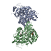
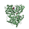


 Mass: 427.201 Da / Num. of mol.: 2 / Source method: obtained synthetically / Formula: C10H15N5O10P2 / Comment: ADP, energy-carrying molecule*YM
 Mass: 24.305 Da / Num. of mol.: 2 / Source method: obtained synthetically / Formula: Mg
 Mass: 58.693 Da / Num. of mol.: 2 / Source method: obtained synthetically / Formula: Ni
 Mass: 59.044 Da / Num. of mol.: 2 / Source method: obtained synthetically / Formula: C2H3O2
 Mass: 92.094 Da / Num. of mol.: 2 / Source method: obtained synthetically / Formula: C3H8O3
 Sample preparation
 / Beamline: PROXIMA 1 / Wavelength: 0.9793
  Processing