 Movie
Movie Controller

[English] 日本語
 Yorodumi
- PDB-1moj: Crystal structure of an archaeal dps-homologue from Halobacterium... -
+ Open data

- Basic information
| Entry | Database: PDB / ID: 1moj | ||||||
|---|---|---|---|---|---|---|---|
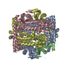
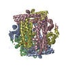
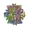
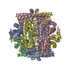
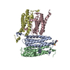
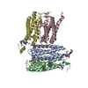
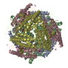
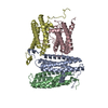
| Title | Crystal structure of an archaeal dps-homologue from Halobacterium salinarum | ||||||
 Components Components | Dps-like ferritin | ||||||
 Keywords Keywords | DNA BINDING PROTEIN / dps / ferritin / four-helix bundle / halophilic | ||||||
| Function / homology |  Function and homology information Function and homology informationOxidoreductases; Oxidizing metal ions / ferric iron binding / intracellular iron ion homeostasis / oxidoreductase activity / cytoplasm Similarity search - Function | ||||||
| Biological species |  Halobacterium salinarum (Halophile) Halobacterium salinarum (Halophile) | ||||||
| Method |  X-RAY DIFFRACTION / SYNCHROTRON / MOLECULAR REPLACEMENT / Resolution: 1.9 Å X-RAY DIFFRACTION / SYNCHROTRON / MOLECULAR REPLACEMENT / Resolution: 1.9 Å | ||||||
 Authors Authors | Zeth, K. / Offermann, S. / Essen, L.O. / Oesterhelt, D. | ||||||
 Citation Citation | Journal: Proc.Natl.Acad.Sci.USA / Year: 2004 Title: Iron-oxo clusters biomineralizing on protein surfaces: Structural analysis of Halobacterium salinarum DpsA in its low- and high-iron states. Authors: Zeth, K. / Offermann, S. / Essen, L.O. / Oesterhelt, D. | ||||||
| History |
|
- Structure visualization
| Structure viewer | Molecule: MolmilJmol/JSmol |
|---|
- Downloads & links
-Download
| PDBx/mmCIF format | 1moj.cif.gz | 150.4 KB | Display | PDBx/mmCIF format |
|---|---|---|---|---|
| PDB format | pdb1moj.ent.gz | 119.2 KB | Display | PDB format |
| PDBx/mmJSON format | 1moj.json.gz | Tree view | PDBx/mmJSON format | |
| Others |  Other downloads Other downloads |
-Validation report
| Arichive directory | https://data.pdbj.org/pub/pdb/validation_reports/mo/1mojftp://data.pdbj.org/pub/pdb/validation_reports/mo/1moj | HTTPS FTP |
|---|
-Related structure data
| Related structure data |  1tjoC  1tk6C  1tkoC  1tkpC 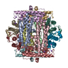 1dpsS S: Starting model for refinement C: citing same article ( |
|---|---|
| Similar structure data |
-Links
 PDBj
PDBj


- Assembly
| Deposited unit | 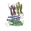
| ||||||||||||
|---|---|---|---|---|---|---|---|---|---|---|---|---|---|
| 1 | 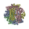
| ||||||||||||
| Unit cell |
| ||||||||||||
| Components on special symmetry positions |
| ||||||||||||
| Details | The biological assembly of a 23-symmetric dodecamer is generated from the tetramer in the asymmetric unit by the operations: -Y,X-Y,Z and Y-X,-X,Z |
-Components
| #1: Protein | Mass: 20121.846 Da / Num. of mol.: 4 / Source method: isolated from a natural source / Source: (natural) Halobacterium salinarum (Halophile) / Strain: TOM / References: UniProt: Q9HMP7#2: Chemical | ChemComp-FE /   Mass: 55.845 Da / Num. of mol.: 6 / Source method: obtained synthetically / Formula: Fe Mass: 55.845 Da / Num. of mol.: 6 / Source method: obtained synthetically / Formula: Fe#3: Chemical |   Mass: 22.990 Da / Num. of mol.: 2 / Source method: obtained synthetically / Formula: Na Mass: 22.990 Da / Num. of mol.: 2 / Source method: obtained synthetically / Formula: Na#4: Chemical | ChemComp-MG /   Mass: 24.305 Da / Num. of mol.: 4 / Source method: obtained synthetically / Formula: Mg Mass: 24.305 Da / Num. of mol.: 4 / Source method: obtained synthetically / Formula: Mg#5: Water | ChemComp-HOH / |  Mass: 18.015 Da / Num. of mol.: 297 / Source method: isolated from a natural source / Formula: H2O Mass: 18.015 Da / Num. of mol.: 297 / Source method: isolated from a natural source / Formula: H2O |
|---|
-Experimental details
-Experiment
| Experiment | Method: X-RAY DIFFRACTION / Number of used crystals: 1 |
|---|
- Sample preparation
| Crystal | Density Matthews: 2.21 Å3/Da / Density % sol: 44.25 % |
|---|---|
| Crystal grow | Temperature: 295 K / Method: vapor diffusion, hanging drop / pH: 8 Details: PEG 400, Tris/HCl, magnesium chloride, sodium chloride, pH 8.0, VAPOR DIFFUSION, HANGING DROP, temperature 295K |
-Data collection
| Diffraction | Mean temperature: 100 K |
|---|---|
| Diffraction source | Source: SYNCHROTRON / Site: ESRF  / Beamline: BM14 / Wavelength: 0.9762 Å / Beamline: BM14 / Wavelength: 0.9762 Å |
| Detector | Type: MAR CCD 165 mm / Detector: CCD / Date: Jun 16, 2001 / Details: mirrors |
| Radiation | Monochromator: mirrors / Protocol: SINGLE WAVELENGTH / Monochromatic (M) / Laue (L): M / Scattering type: x-ray |
| Radiation wavelength | Wavelength: 0.9762 Å / Relative weight: 1 |
| Reflection | Resolution: 1.8→30 Å / Num. all: 66071 / Num. obs: 63709 / % possible obs: 99.6 % / Observed criterion σ(F): 0 / Observed criterion σ(I): -3 / Redundancy: 5.9 % / Biso Wilson estimate: 13 Å2 / Rmerge(I) obs: 0.096 / Net I/σ(I): 12.3 |
| Reflection shell | Resolution: 1.8→1.99 Å / Rmerge(I) obs: 0.45 / Mean I/σ(I) obs: 4.1 / % possible all: 99.3 |
- Processing
| Software |
| ||||||||||||||||||||||||||||||||||||
|---|---|---|---|---|---|---|---|---|---|---|---|---|---|---|---|---|---|---|---|---|---|---|---|---|---|---|---|---|---|---|---|---|---|---|---|---|---|
| Refinement | Method to determine structure: MOLECULAR REPLACEMENT Starting model: PDB entry 1DPS Resolution: 1.9→29.88 Å / Isotropic thermal model: RESTRAINED / Cross valid method: THROUGHOUT / σ(F): 0 / Stereochemistry target values: Engh & Huber Details: The crystal was merohedrally twinned along the z-axis with a twinning ratio of 0.57/0.43 for relating reflections h,k,l with -h,-k,l. Refinement of the twinned crystal proceeded in CNS, but ...Details: The crystal was merohedrally twinned along the z-axis with a twinning ratio of 0.57/0.43 for relating reflections h,k,l with -h,-k,l. Refinement of the twinned crystal proceeded in CNS, but the twinned R-factors are systematically underestimated compared to the non-twinned case
| ||||||||||||||||||||||||||||||||||||
| Solvent computation | Solvent model: FLAT MODEL / Bsol: 43.7172 Å2 / ksol: 0.427365 e/Å3 | ||||||||||||||||||||||||||||||||||||
| Displacement parameters | Biso mean: 18.5 Å2
| ||||||||||||||||||||||||||||||||||||
| Refinement step | Cycle: LAST / Resolution: 1.9→29.88 Å
| ||||||||||||||||||||||||||||||||||||
| Refine LS restraints |
| ||||||||||||||||||||||||||||||||||||
| Refine LS restraints NCS | NCS model details: CONSTR | ||||||||||||||||||||||||||||||||||||
| LS refinement shell | Resolution: 1.9→1.99 Å / Total num. of bins used: 8
| ||||||||||||||||||||||||||||||||||||
| Xplor file |
|
