 Movie
Movie Controller


+ Open data

- Basic information
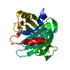
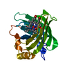
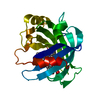
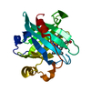
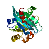
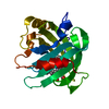
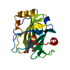
| Entry | Database: PDB / ID: 1ml7 | ||||||
|---|---|---|---|---|---|---|---|
| Title | Crystal structure of nitrophorin 4 complexed with 4-iodopyrazole | ||||||
 Components Components | nitrophorin 4 | ||||||
 Keywords Keywords | LIGAND BINDING PROTEIN / NO carrier / ferric heme / iodopyrazole / lipocalin / beta barrel / conformational change | ||||||
| Function / homology |  Function and homology information Function and homology informationnitrite dismutase / histamine binding / nitric oxide binding / vasodilation / oxidoreductase activity / extracellular region / metal ion binding Similarity search - Function | ||||||
| Biological species |   Rhodnius prolixus (insect) Rhodnius prolixus (insect) | ||||||
| Method |  X-RAY DIFFRACTION / SYNCHROTRON / FOURIER SYNTHESIS / Resolution: 1.25 Å X-RAY DIFFRACTION / SYNCHROTRON / FOURIER SYNTHESIS / Resolution: 1.25 Å | ||||||
 Authors Authors | Berry, R.E. / Ding, X.D. / Weichsel, A. / Montfort, W.R. / Walker, F.A. | ||||||
 Citation Citation | Journal: J.Biol.Inorg.Chem. / Year: 2004 Title: Axial ligand complexes of the Rhodnius nitrophorins: reduction potentials, binding constants, EPR spectra, and structures of the 4-iodopyrazole and imidazole complexes of NP4 Authors: Berry, R.E. / Ding, X.D. / Shokhireva, T.K. / Weichsel, A. / Montfort, W.R. / Walker, F.A. #1: Journal: Biochemistry / Year: 2001Title: Ligand-Induced Heme Ruffling and Bent NO Geometry in Ultra-High-Resolution Structures of Nitophorin 4 Authors: Roberts, S.A. / Weichsel, A. / Qiu, Y. / Shelnutt, J.A. / Walker, F.A. / Montfort, W.R. | ||||||
| History |
|
- Structure visualization
| Structure viewer | Molecule: MolmilJmol/JSmol |
|---|
- Downloads & links
-Download
| PDBx/mmCIF format | 1ml7.cif.gz | 97.9 KB | Display | PDBx/mmCIF format |
|---|---|---|---|---|
| PDB format | pdb1ml7.ent.gz | 73.7 KB | Display | PDB format |
| PDBx/mmJSON format | 1ml7.json.gz | Tree view | PDBx/mmJSON format | |
| Others |  Other downloads Other downloads |
-Validation report
| Summary document | 1ml7_validation.pdf.gz | 794 KB | Display | wwPDB validaton report |
|---|---|---|---|---|
| Full document | 1ml7_full_validation.pdf.gz | 796.2 KB | Display | |
| Data in XML | 1ml7_validation.xml.gz | 14.3 KB | Display | |
| Data in CIF | 1ml7_validation.cif.gz | 20.2 KB | Display | |
| Arichive directory | https://data.pdbj.org/pub/pdb/validation_reports/ml/1ml7ftp://data.pdbj.org/pub/pdb/validation_reports/ml/1ml7 | HTTPS FTP |
-Related structure data
| Related structure data |  1ikjS S: Starting model for refinement |
|---|---|
| Similar structure data |
-Links
 PDBj
PDBj


- Assembly
| Deposited unit | 
| |||||||||||||||||||||
|---|---|---|---|---|---|---|---|---|---|---|---|---|---|---|---|---|---|---|---|---|---|---|
| 1 |
| |||||||||||||||||||||
| Unit cell |
| |||||||||||||||||||||
| Components on special symmetry positions |
| |||||||||||||||||||||
| Details | The biological assembly is a monomer |
-Components
| #1: Protein | Mass: 20292.664 Da / Num. of mol.: 1 Source method: isolated from a genetically manipulated source Source: (gene. exp.) Rhodnius prolixus (insect) / Production host:  | ||||
|---|---|---|---|---|---|
| #2: Chemical | ChemComp-HEV / 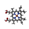  Mass: 640.509 Da / Num. of mol.: 1 / Source method: obtained synthetically / Formula: C36H32FeN4O4 Mass: 640.509 Da / Num. of mol.: 1 / Source method: obtained synthetically / Formula: C36H32FeN4O4 | ||||
| #3: Chemical |   Mass: 193.974 Da / Num. of mol.: 2 / Source method: obtained synthetically / Formula: C3H3IN2 Mass: 193.974 Da / Num. of mol.: 2 / Source method: obtained synthetically / Formula: C3H3IN2#4: Water | ChemComp-HOH / |  Mass: 18.015 Da / Num. of mol.: 244 / Source method: isolated from a natural source / Formula: H2O Mass: 18.015 Da / Num. of mol.: 244 / Source method: isolated from a natural source / Formula: H2OHas protein modification | Y | |
-Experimental details
-Experiment
| Experiment | Method: X-RAY DIFFRACTION / Number of used crystals: 1 |
|---|
- Sample preparation
| Crystal | Density Matthews: 1.95 Å3/Da / Density % sol: 36.99 % | ||||||||||||||||||
|---|---|---|---|---|---|---|---|---|---|---|---|---|---|---|---|---|---|---|---|
| Crystal grow | Temperature: 298 K / Method: vapor diffusion, hanging drop / pH: 7.5 Details: ammonium phosphate, pH 7.5, VAPOR DIFFUSION, HANGING DROP at 298K, temperature 298.0K | ||||||||||||||||||
| Crystal grow | *PLUS | ||||||||||||||||||
| Components of the solutions | *PLUS
|
-Data collection
| Diffraction | Mean temperature: 100 K |
|---|---|
| Diffraction source | Source: SYNCHROTRON / Site: SSRL  / Beamline: BL7-1 / Wavelength: 1.08 Å / Beamline: BL7-1 / Wavelength: 1.08 Å |
| Detector | Type: MARRESEARCH / Detector: IMAGE PLATE / Date: May 17, 2002 |
| Radiation | Monochromator: Cyclindrically bent triangular Si(111) asymmetric cut Protocol: SINGLE WAVELENGTH / Monochromatic (M) / Laue (L): M / Scattering type: x-ray |
| Radiation wavelength | Wavelength: 1.08 Å / Relative weight: 1 |
| Reflection | Resolution: 1.25→22 Å / Num. all: 42458 / Num. obs: 42458 / % possible obs: 97.8 % / Observed criterion σ(F): 0 / Observed criterion σ(I): 0 / Redundancy: 5.1 % / Biso Wilson estimate: 7.85 Å2 / Rmerge(I) obs: 0.05 / Net I/σ(I): 21.7 |
| Reflection shell | Resolution: 1.25→1.29 Å / Redundancy: 4.7 % / Rmerge(I) obs: 0.158 / Mean I/σ(I) obs: 7.2 / Num. unique all: 3947 / % possible all: 92 |
| Reflection | *PLUS Lowest resolution: 21 Å / % possible obs: 98 % / Num. measured all: 219394 |
| Reflection shell | *PLUS % possible obs: 92 % / Rmerge(I) obs: 0.16 |
- Processing
| Software |
| |||||||||||||||||||||||||
|---|---|---|---|---|---|---|---|---|---|---|---|---|---|---|---|---|---|---|---|---|---|---|---|---|---|---|
| Refinement | Method to determine structure: FOURIER SYNTHESIS Starting model: PDB entry 1IKJ Resolution: 1.25→20.4 Å / Isotropic thermal model: anisotropic / Cross valid method: THROUGHOUT / σ(F): 0 / Stereochemistry target values: Engh & Huber
| |||||||||||||||||||||||||
| Displacement parameters | Biso mean: 12.8 Å2 | |||||||||||||||||||||||||
| Refinement step | Cycle: LAST / Resolution: 1.25→20.4 Å
| |||||||||||||||||||||||||
| Refine LS restraints |
| |||||||||||||||||||||||||
| Software | *PLUS Name: SHELXL / Version: 97 / Classification: refinement | |||||||||||||||||||||||||
| Refinement | *PLUS % reflection Rfree: 5 % | |||||||||||||||||||||||||
| Solvent computation | *PLUS | |||||||||||||||||||||||||
| Displacement parameters | *PLUS | |||||||||||||||||||||||||
| Refine LS restraints | *PLUS
|
