 Movie
Movie Controller

[English] 日本語
 Yorodumi
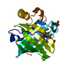
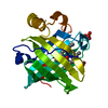
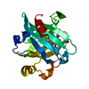
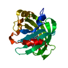
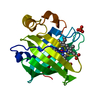
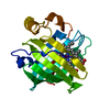
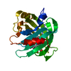
- PDB-1ikj: 1.27 A CRYSTAL STRUCTURE OF NITROPHORIN 4 FROM RHODNIUS PROLIXUS ... -
+ Open data

- Basic information
| Entry | Database: PDB / ID: 1ikj | ||||||
|---|---|---|---|---|---|---|---|
| Title | 1.27 A CRYSTAL STRUCTURE OF NITROPHORIN 4 FROM RHODNIUS PROLIXUS COMPLEXED WITH IMIDAZOLE | ||||||
 Components Components | NITROPHORIN 4 | ||||||
 Keywords Keywords | TRANSPORT PROTEIN / NITRIC OXIDE TRANSPORT / FERRIC HEME / ANTIHISTAMINE / VASODILATOR / LIPOCALIN / BILAN BINDING PROTEIN | ||||||
| Function / homology |  Function and homology information Function and homology informationnitrite dismutase / histamine binding / nitric oxide binding / vasodilation / oxidoreductase activity / extracellular region / metal ion binding Similarity search - Function | ||||||
| Biological species |  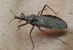 Rhodnius prolixus (insect) Rhodnius prolixus (insect) | ||||||
| Method |  X-RAY DIFFRACTION / Resolution: 1.27 Å X-RAY DIFFRACTION / Resolution: 1.27 Å | ||||||
 Authors Authors | Roberts, S.A. / Weichsel, A. / Qui, Y. / Shelnutt, J.A. / Walker, F.A. / Montfort, W.R. | ||||||
 Citation Citation | Journal: Biochemistry / Year: 2001 Title: Ligand-induced heme ruffling and bent no geometry in ultra-high-resolution structures of nitrophorin 4. Authors: Roberts, S.A. / Weichsel, A. / Qiu, Y. / Shelnutt, J.A. / Walker, F.A. / Montfort, W.R. | ||||||
| History |
|
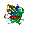
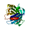
- Structure visualization
| Structure viewer | Molecule: MolmilJmol/JSmol |
|---|
- Downloads & links
-Download
| PDBx/mmCIF format | 1ikj.cif.gz | 102.4 KB | Display | PDBx/mmCIF format |
|---|---|---|---|---|
| PDB format | pdb1ikj.ent.gz | 77.7 KB | Display | PDB format |
| PDBx/mmJSON format | 1ikj.json.gz | Tree view | PDBx/mmJSON format | |
| Others |  Other downloads Other downloads |
-Validation report
| Arichive directory | https://data.pdbj.org/pub/pdb/validation_reports/ik/1ikjftp://data.pdbj.org/pub/pdb/validation_reports/ik/1ikj | HTTPS FTP |
|---|
-Related structure data
| Related structure data | 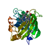 1d2uC  1ikeC  1koiC  1ikh C: citing same article ( S: Starting model for refinement |
|---|---|
| Similar structure data |


-Links
 PDBj
PDBj


- Assembly
| Deposited unit | 
| ||||||||||||
|---|---|---|---|---|---|---|---|---|---|---|---|---|---|
| 1 |
| ||||||||||||
| Unit cell |
| ||||||||||||
| Components on special symmetry positions |
|
-Components
| #1: Protein | Mass: 20292.664 Da / Num. of mol.: 1 Source method: isolated from a genetically manipulated source Details: IMIDAZOLE OCCUPIES THE FE SIXTH COORDINATION POSITION THE HEME IS DISORDERED BY A ROTATION OF 180 DEGREES AROUND THE CHA-FE-CHC AXIS. THE ONLY EVIDENCE OF THIS DISORDER IS THE APPEARANCE OF ...Details: IMIDAZOLE OCCUPIES THE FE SIXTH COORDINATION POSITION THE HEME IS DISORDERED BY A ROTATION OF 180 DEGREES AROUND THE CHA-FE-CHC AXIS. THE ONLY EVIDENCE OF THIS DISORDER IS THE APPEARANCE OF METHYL GROUPS CMB AND CMC AS VINYLS. THESE EXTRA ATOMS ARE CALLED CBBB AND CBCB. THE LOOP CONTAINING RESIDUES 125-130 IS DISORDERED. IT HAS BEEN BUILT IN TWO POSITIONS, BUT THIS MODEL DOES NOT COMPLETELY EXPLAIN THE ELECTRON DENSITY. Source: (gene. exp.) Rhodnius prolixus (insect) / Plasmid: PET-17B-NP4 / Species (production host): Escherichia coli / Production host:  |
|---|---|
| #2: Chemical | ChemComp-HEM /   Mass: 616.487 Da / Num. of mol.: 1 / Source method: obtained synthetically / Formula: C34H32FeN4O4 Mass: 616.487 Da / Num. of mol.: 1 / Source method: obtained synthetically / Formula: C34H32FeN4O4 |
| #3: Chemical | ChemComp-IMD /   Mass: 69.085 Da / Num. of mol.: 1 / Source method: obtained synthetically / Formula: C3H5N2 Mass: 69.085 Da / Num. of mol.: 1 / Source method: obtained synthetically / Formula: C3H5N2 |
| #4: Water | ChemComp-HOH /  Mass: 18.015 Da / Num. of mol.: 276 / Source method: isolated from a natural source / Formula: H2O Mass: 18.015 Da / Num. of mol.: 276 / Source method: isolated from a natural source / Formula: H2O |
| Has protein modification | Y |
-Experimental details
-Experiment
| Experiment | Method: X-RAY DIFFRACTION / Number of used crystals: 1 |
|---|
- Sample preparation
| Crystal | Density Matthews: 1.94 Å3/Da / Density % sol: 36.58 % |
|---|---|
| Crystal grow | Temperature: 295 K / Method: vapor diffusion, hanging drop / pH: 5.6 Details: PEG-4000, sodium citrate, pH 5.60, VAPOR DIFFUSION, HANGING DROP, temperature 295K |
-Data collection
| Diffraction | Mean temperature: 100 K |
|---|---|
| Diffraction source | Source: ROTATING ANODE / Type: RIGAKU RUH3R / Wavelength: 1.5418 / Wavelength: 1.5418 Å |
| Detector | Type: RIGAKU RAXIS IV++ / Detector: IMAGE PLATE / Date: Feb 2, 2001 / Details: MSC Blue Confocal Mirrors |
| Radiation | Monochromator: mirrors / Protocol: SINGLE WAVELENGTH / Monochromatic (M) / Laue (L): M / Scattering type: x-ray |
| Radiation wavelength | Wavelength: 1.5418 Å / Relative weight: 1 |
| Reflection | Resolution: 1.22→22 Å / Num. all: 40637 / Num. obs: 37957 / % possible obs: 96 % / Observed criterion σ(F): 0 / Observed criterion σ(I): 0 / Redundancy: 2.4 % / Biso Wilson estimate: 9.1 Å2 / Rmerge(I) obs: 0.032 / Net I/σ(I): 14 |
| Reflection shell | Resolution: 1.27→1.3 Å / Redundancy: 1.4 % / Rmerge(I) obs: 0.065 / Mean I/σ(I) obs: 9.2 / % possible all: 79 |
- Processing
| Software |
| |||||||||||||||||||||||||||||||||
|---|---|---|---|---|---|---|---|---|---|---|---|---|---|---|---|---|---|---|---|---|---|---|---|---|---|---|---|---|---|---|---|---|---|---|
| Refinement | Starting model: pdb entry 1IKH 1ikh Resolution: 1.27→22 Å / Num. parameters: 16345 / Num. restraintsaints: 19521 Isotropic thermal model: Anisotropic temperature factors for all non-hydrogen atoms σ(F): 0 / σ(I): 0 / Stereochemistry target values: ENGH & HUBER Details: conjugate gradient refinement with SHELX, all atoms refined with anisotropic thermal parameters, hydrogen atoms added at calculated positions. Block-diagonal matrix refinement of the heme ...Details: conjugate gradient refinement with SHELX, all atoms refined with anisotropic thermal parameters, hydrogen atoms added at calculated positions. Block-diagonal matrix refinement of the heme and iron coordination sphere without restraints.
| |||||||||||||||||||||||||||||||||
| Refine analyze | Num. disordered residues: 16 | |||||||||||||||||||||||||||||||||
| Refinement step | Cycle: LAST / Resolution: 1.27→22 Å
| |||||||||||||||||||||||||||||||||
| Refine LS restraints |
|
