 Movie
Movie Controller

[English] 日本語
 Yorodumi
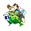
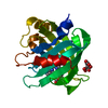
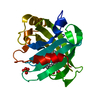
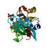
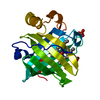
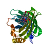
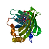
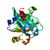
- PDB-1koi: CRYSTAL STRUCTURE OF NITROPHORIN 4 FROM RHODNIUS PROLIXUS COMPLEX... -
+ Open data

- Basic information
| Entry | Database: PDB / ID: 1koi | |||||||||
|---|---|---|---|---|---|---|---|---|---|---|
| Title | CRYSTAL STRUCTURE OF NITROPHORIN 4 FROM RHODNIUS PROLIXUS COMPLEXED WITH NITRIC OXIDE AT 1.08 A RESOLUTION | |||||||||
 Components Components | NITROPHORIN 4 | |||||||||
 Keywords Keywords | TRANSPORT PROTEIN / nitric oxide transport / ferric heme / anithistimine / lipocalin | |||||||||
| Function / homology |  Function and homology information Function and homology informationnitrite dismutase / histamine binding / nitric oxide binding / vasodilation / oxidoreductase activity / extracellular region / metal ion binding Similarity search - Function | |||||||||
| Biological species |  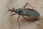 Rhodnius prolixus (insect) Rhodnius prolixus (insect) | |||||||||
| Method |  X-RAY DIFFRACTION / SYNCHROTRON / Resolution: 1.08 Å X-RAY DIFFRACTION / SYNCHROTRON / Resolution: 1.08 Å | |||||||||
 Authors Authors | Roberts, S.A. / Weichsel, A. / Qiu, Y. / Shelnutt, J.A. / Walker, F.A. / Montfort, W.R. | |||||||||
 Citation Citation | Journal: Biochemistry / Year: 2001 Title: Ligand-induced heme ruffling and bent no geometry in ultra-high-resolution structures of nitrophorin 4. Authors: Roberts, S.A. / Weichsel, A. / Qiu, Y. / Shelnutt, J.A. / Walker, F.A. / Montfort, W.R. | |||||||||
| History |
|
- Structure visualization
| Structure viewer | Molecule: MolmilJmol/JSmol |
|---|
- Downloads & links
-Download
| PDBx/mmCIF format | 1koi.cif.gz | 98.7 KB | Display | PDBx/mmCIF format |
|---|---|---|---|---|
| PDB format | pdb1koi.ent.gz | 74.3 KB | Display | PDB format |
| PDBx/mmJSON format | 1koi.json.gz | Tree view | PDBx/mmJSON format | |
| Others |  Other downloads Other downloads |
-Validation report
| Arichive directory | https://data.pdbj.org/pub/pdb/validation_reports/ko/1koiftp://data.pdbj.org/pub/pdb/validation_reports/ko/1koi | HTTPS FTP |
|---|
-Related structure data
| Related structure data | 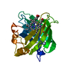 1d2uC  1ikeC  1ikjC  1erxS S: Starting model for refinement C: citing same article ( |
|---|---|
| Similar structure data |

-Links
 PDBj
PDBj


- Assembly
| Deposited unit | 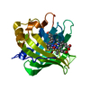
| ||||||||||||
|---|---|---|---|---|---|---|---|---|---|---|---|---|---|
| 1 |
| ||||||||||||
| Unit cell |
| ||||||||||||
| Components on special symmetry positions |
|
-Components
| #1: Protein | Mass: 20292.664 Da / Num. of mol.: 1 Source method: isolated from a genetically manipulated source Details: NITRIC OXIDE OCCUPIES THE SIXTH COORDINATION POSITION OF THE IRON. THE HEME IS DISORDERED BY A ROTATION OF 180 DEGREES AROUND THE CHA-FE-CHC AXIS. THE ONLY EVIDENCE OF THIS DISORDER IS THE ...Details: NITRIC OXIDE OCCUPIES THE SIXTH COORDINATION POSITION OF THE IRON. THE HEME IS DISORDERED BY A ROTATION OF 180 DEGREES AROUND THE CHA-FE-CHC AXIS. THE ONLY EVIDENCE OF THIS DISORDER IS THE APPEARANCE OF METHYL GROUPS CMB AND CMC AS VINYLS. THESE EXTRA ATOMS ARE CALLED CBBB AND CBCB. Source: (gene. exp.) Rhodnius prolixus (insect) / Plasmid: PET-17B-NP4 / Species (production host): Escherichia coli / Production host:  |
|---|---|
| #2: Chemical | ChemComp-HEM /   Mass: 616.487 Da / Num. of mol.: 1 / Source method: obtained synthetically / Formula: C34H32FeN4O4 Mass: 616.487 Da / Num. of mol.: 1 / Source method: obtained synthetically / Formula: C34H32FeN4O4 |
| #3: Chemical | ChemComp-NO /   Mass: 30.006 Da / Num. of mol.: 1 / Source method: obtained synthetically / Formula: NO Mass: 30.006 Da / Num. of mol.: 1 / Source method: obtained synthetically / Formula: NO |
| #4: Water | ChemComp-HOH /  Mass: 18.015 Da / Num. of mol.: 264 / Source method: isolated from a natural source / Formula: H2O Mass: 18.015 Da / Num. of mol.: 264 / Source method: isolated from a natural source / Formula: H2O |
| Has protein modification | Y |
-Experimental details
-Experiment
| Experiment | Method: X-RAY DIFFRACTION / Number of used crystals: 1 |
|---|
- Sample preparation
| Crystal | Density Matthews: 1.94 Å3/Da / Density % sol: 36.44 % |
|---|---|
| Crystal grow | Temperature: 295 K / Method: vapor diffusion, hanging drop / pH: 5.6 Details: PEG 4000, Sodium Citrate, pH 5.6, VAPOR DIFFUSION, HANGING DROP, temperature 295K |
-Data collection
| Diffraction | Mean temperature: 100 K |
|---|---|
| Diffraction source | Source: SYNCHROTRON / Site: NSLS  / Beamline: X12B / Wavelength: 0.975 Å / Beamline: X12B / Wavelength: 0.975 Å |
| Detector | Type: ADSC QUANTUM 4 / Detector: CCD / Date: Mar 1, 2000 |
| Radiation | Protocol: SINGLE WAVELENGTH / Monochromatic (M) / Laue (L): M / Scattering type: x-ray |
| Radiation wavelength | Wavelength: 0.975 Å / Relative weight: 1 |
| Reflection | Resolution: 1→30 Å / Num. all: 83813 / Num. obs: 62726 / % possible obs: 95 % / Observed criterion σ(F): 0 / Observed criterion σ(I): -3 / Redundancy: 5 % / Biso Wilson estimate: 4.3 Å2 / Rmerge(I) obs: 0.04 / Net I/σ(I): 38 |
| Reflection shell | Resolution: 1.08→1.1 Å / Redundancy: 2.4 % / Rmerge(I) obs: 0.24 / Mean I/σ(I) obs: 15 / % possible all: 70 |
- Processing
| Software |
| ||||||||||||||||||||
|---|---|---|---|---|---|---|---|---|---|---|---|---|---|---|---|---|---|---|---|---|---|
| Refinement | Starting model: pdb entry 1ERX Resolution: 1.08→30 Å / Isotropic thermal model: All non-hydrogen atoms anisotropic / Cross valid method: THROUGHOUT / σ(F): 0 / σ(I): 0 / Stereochemistry target values: Engh & Huber Details: Shelx conjugate gradient refinement. Hydrogen atoms added at calculated positions. Heme and iron coordination sphere refined without restraints.
| ||||||||||||||||||||
| Displacement parameters | Biso mean: 13 Å2 | ||||||||||||||||||||
| Refinement step | Cycle: LAST / Resolution: 1.08→30 Å
|
