 Movie
Movie Controller

[English] 日本語
 Yorodumi
- PDB-1ml5: Structure of the E. coli ribosomal termination complex with relea... -
+ Open data

- Basic information
| Entry | Database: PDB / ID: 1ml5 | ||||||
|---|---|---|---|---|---|---|---|
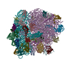
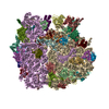
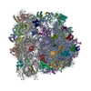
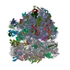
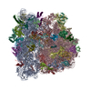
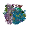
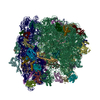
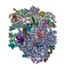
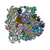
| Title | Structure of the E. coli ribosomal termination complex with release factor 2 | ||||||
 Components Components |
| ||||||
 Keywords Keywords | RIBOSOME / E. coli / termination of protein synthesis / release factor / cryo-eletron microscopy / angular reconstitution | ||||||
| Function / homology |  Function and homology information Function and homology informationtranslation release factor activity, codon specific / translational termination / response to radiation / regulation of translation / large ribosomal subunit / transferase activity / ribosomal small subunit assembly / ribosomal small subunit biogenesis / 5S rRNA binding / ribosomal large subunit assembly ...translation release factor activity, codon specific / translational termination / response to radiation / regulation of translation / large ribosomal subunit / transferase activity / ribosomal small subunit assembly / ribosomal small subunit biogenesis / 5S rRNA binding / ribosomal large subunit assembly / small ribosomal subunit / small ribosomal subunit rRNA binding / large ribosomal subunit rRNA binding / cytosolic small ribosomal subunit / cytosolic large ribosomal subunit / cytoplasmic translation / tRNA binding / negative regulation of translation / rRNA binding / structural constituent of ribosome / ribosome / translation / ribonucleoprotein complex / viral translational frameshifting / mRNA binding / zinc ion binding / metal ion binding / cytoplasm / cytosol Similarity search - Function | ||||||
| Biological species |  | ||||||
| Method | ELECTRON MICROSCOPY / single particle reconstruction / cryo EM / Resolution: 14 Å | ||||||
 Authors Authors | Klaholz, B.P. / Pape, T. / Zavialov, A.V. / Myasnikov, A.G. / Orlova, E.V. / Vestergaard, B. / Ehrenberg, M. / van Heel, M. | ||||||
 Citation Citation | Journal: Nature / Year: 2003 Title: Structure of the Escherichia coli ribosomal termination complex with release factor 2. Authors: Bruno P Klaholz / Tillmann Pape / Andrey V Zavialov / Alexander G Myasnikov / Elena V Orlova / Bente Vestergaard / Måns Ehrenberg / Marin van Heel /  Abstract: Termination of protein synthesis occurs when the messenger RNA presents a stop codon in the ribosomal aminoacyl (A) site. Class I release factor proteins (RF1 or RF2) are believed to recognize stop ...Termination of protein synthesis occurs when the messenger RNA presents a stop codon in the ribosomal aminoacyl (A) site. Class I release factor proteins (RF1 or RF2) are believed to recognize stop codons via tripeptide motifs, leading to release of the completed polypeptide chain from its covalent attachment to transfer RNA in the ribosomal peptidyl (P) site. Class I RFs possess a conserved GGQ amino-acid motif that is thought to be involved directly in protein-transfer-RNA bond hydrolysis. Crystal structures of bacterial and eukaryotic class I RFs have been determined, but the mechanism of stop codon recognition and peptidyl-tRNA hydrolysis remains unclear. Here we present the structure of the Escherichia coli ribosome in a post-termination complex with RF2, obtained by single-particle cryo-electron microscopy (cryo-EM). Fitting the known 70S and RF2 structures into the electron density map reveals that RF2 adopts a different conformation on the ribosome when compared with the crystal structure of the isolated protein. The amino-terminal helical domain of RF2 contacts the factor-binding site of the ribosome, the 'SPF' loop of the protein is situated close to the mRNA, and the GGQ-containing domain of RF2 interacts with the peptidyl-transferase centre (PTC). By connecting the ribosomal decoding centre with the PTC, RF2 functionally mimics a tRNA molecule in the A site. Translational termination in eukaryotes is likely to be based on a similar mechanism. | ||||||
| History |
|
- Structure visualization
| Movie |
Movie viewer |
|---|---|
| Structure viewer | Molecule: MolmilJmol/JSmol |
- Downloads & links
-Download
| PDBx/mmCIF format | 1ml5.cif.gz | 398 KB | Display | PDBx/mmCIF format |
|---|---|---|---|---|
| PDB format | pdb1ml5.ent.gz | 222.6 KB | Display | PDB format |
| PDBx/mmJSON format | 1ml5.json.gz | Tree view | PDBx/mmJSON format | |
| Others |  Other downloads Other downloads |
-Validation report
| Arichive directory | https://data.pdbj.org/pub/pdb/validation_reports/ml/1ml5ftp://data.pdbj.org/pub/pdb/validation_reports/ml/1ml5 | HTTPS FTP |
|---|
-Related structure data
| Related structure data |  1005MC M: map data used to model this data C: citing same article ( |
|---|---|
| Similar structure data |


-Links
 PDBj
PDBj


- Assembly
| Deposited unit | 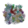
|
|---|---|
| 1 |
|
-Components
-RNA chain , 5 types, 5 molecules ABCab
| #1: RNA chain | Mass: 493958.281 Da / Num. of mol.: 1 / Source method: isolated from a natural source / Source: (natural) |
|---|---|
| #2: RNA chain | Mass: 24890.121 Da / Num. of mol.: 1 / Source method: isolated from a natural source / Details: tRNA P-SITE / Source: (natural) |
| #3: RNA chain | Mass: 1792.037 Da / Num. of mol.: 1 / Source method: isolated from a natural source / Details: 6 NT LONG MRNA FRAGMENT / Source: (natural) |
| #4: RNA chain | Mass: 948280.688 Da / Num. of mol.: 1 / Source method: isolated from a natural source / Source: (natural) |
| #5: RNA chain | Mass: 39846.781 Da / Num. of mol.: 1 / Source method: isolated from a natural source / Source: (natural) |
-Protein , 1 types, 1 molecules Z
| #6: Protein | Mass: 41300.660 Da / Num. of mol.: 1 Source method: isolated from a genetically manipulated source Details: EM reconstruction of the backbone trace of the ribosome Source: (gene. exp.) |
|---|
-30S RIBOSOMAL PROTEIN ... , 20 types, 20 molecules EFGHIJKLMNOPQRSTUVWX
| #7: Protein | Mass: 29317.703 Da / Num. of mol.: 1 / Source method: isolated from a natural source / Source: (natural) |
|---|---|
| #8: Protein | Mass: 26751.076 Da / Num. of mol.: 1 / Source method: isolated from a natural source / Source: (natural) |
| #9: Protein | Mass: 24373.447 Da / Num. of mol.: 1 / Source method: isolated from a natural source / Source: (natural) |
| #10: Protein | Mass: 17583.416 Da / Num. of mol.: 1 / Source method: isolated from a natural source / Source: (natural) |
| #11: Protein | Mass: 11988.753 Da / Num. of mol.: 1 / Source method: isolated from a natural source / Source: (natural) |
| #12: Protein | Mass: 18050.973 Da / Num. of mol.: 1 / Source method: isolated from a natural source / Source: (natural) |
| #13: Protein | Mass: 15868.570 Da / Num. of mol.: 1 / Source method: isolated from a natural source / Source: (natural) |
| #14: Protein | Mass: 14429.661 Da / Num. of mol.: 1 / Source method: isolated from a natural source / Source: (natural) |
| #15: Protein | Mass: 11954.968 Da / Num. of mol.: 1 / Source method: isolated from a natural source / Source: (natural) |
| #16: Protein | Mass: 13737.868 Da / Num. of mol.: 1 / Source method: isolated from a natural source / Source: (natural) |
| #17: Protein | Mass: 14920.754 Da / Num. of mol.: 1 / Source method: isolated from a natural source / Source: (natural) |
| #18: Protein | Mass: 14338.861 Da / Num. of mol.: 1 / Source method: isolated from a natural source / Source: (natural) |
| #19: Protein | Mass: 7158.725 Da / Num. of mol.: 1 / Source method: isolated from a natural source / Source: (natural) |
| #20: Protein | Mass: 10578.407 Da / Num. of mol.: 1 / Source method: isolated from a natural source / Source: (natural) |
| #21: Protein | Mass: 10808.419 Da / Num. of mol.: 1 / Source method: isolated from a natural source / Source: (natural) |
| #22: Protein | Mass: 12324.670 Da / Num. of mol.: 1 / Source method: isolated from a natural source / Source: (natural) |
| #23: Protein | Mass: 10244.272 Da / Num. of mol.: 1 / Source method: isolated from a natural source / Source: (natural) |
| #24: Protein | Mass: 10605.464 Da / Num. of mol.: 1 / Source method: isolated from a natural source / Source: (natural) |
| #25: Protein | Mass: 11722.116 Da / Num. of mol.: 1 / Source method: isolated from a natural source / Source: (natural) |
| #26: Protein/peptide | Mass: 3218.835 Da / Num. of mol.: 1 / Source method: isolated from a natural source / Source: (natural) |
-50S RIBOSOMAL PROTEIN ... , 19 types, 19 molecules cdefghlmnopqrstuvwx
| #27: Protein | Mass: 24736.502 Da / Num. of mol.: 1 / Source method: isolated from a natural source / Source: (natural) |
|---|---|
| #28: Protein | Mass: 19283.371 Da / Num. of mol.: 1 / Source method: isolated from a natural source / Source: (natural) |
| #29: Protein | Mass: 37412.496 Da / Num. of mol.: 1 / Source method: isolated from a natural source / Source: (natural) |
| #30: Protein | Mass: 26443.082 Da / Num. of mol.: 1 / Source method: isolated from a natural source / Source: (natural) |
| #31: Protein | Mass: 19420.498 Da / Num. of mol.: 1 / Source method: isolated from a natural source / Source: (natural) |
| #32: Protein | Mass: 19202.123 Da / Num. of mol.: 1 / Source method: isolated from a natural source / Source: (natural) |
| #33: Protein | Mass: 15111.923 Da / Num. of mol.: 1 / Source method: isolated from a natural source / Source: (natural) |
| #34: Protein | Mass: 16249.214 Da / Num. of mol.: 1 / Source method: isolated from a natural source / Source: (natural) |
| #35: Protein | Mass: 13369.613 Da / Num. of mol.: 1 / Source method: isolated from a natural source / Source: (natural) |
| #36: Protein | Mass: 17874.172 Da / Num. of mol.: 1 / Source method: isolated from a natural source / Source: (natural) |
| #37: Protein | Mass: 15313.307 Da / Num. of mol.: 1 / Source method: isolated from a natural source / Source: (natural) |
| #38: Protein | Mass: 20509.740 Da / Num. of mol.: 1 / Source method: isolated from a natural source / Source: (natural) |
| #39: Protein | Mass: 7233.720 Da / Num. of mol.: 1 / Source method: isolated from a natural source / Source: (natural) |
| #40: Protein | Mass: 12808.055 Da / Num. of mol.: 1 / Source method: isolated from a natural source / Source: (natural) |
| #41: Protein | Mass: 9481.340 Da / Num. of mol.: 1 / Source method: isolated from a natural source / Source: (natural) |
| #42: Protein | Mass: 13539.759 Da / Num. of mol.: 1 / Source method: isolated from a natural source / Source: (natural) |
| #43: Protein | Mass: 10713.465 Da / Num. of mol.: 1 / Source method: isolated from a natural source / Source: (natural) |
| #44: Protein | Mass: 7758.667 Da / Num. of mol.: 1 / Source method: isolated from a natural source / Source: (natural) |
| #45: Protein | Mass: 6799.126 Da / Num. of mol.: 1 / Source method: isolated from a natural source / Source: (natural) |
-Experimental details
-Experiment
| Experiment | Method: ELECTRON MICROSCOPY |
|---|---|
| EM experiment | Aggregation state: PARTICLE / 3D reconstruction method: single particle reconstruction |
- Sample preparation
| Component |
| |||||||||||||||
|---|---|---|---|---|---|---|---|---|---|---|---|---|---|---|---|---|
| Buffer solution | Name: polymix buffer / pH: 7.5 / Details: polymix buffer | |||||||||||||||
| Specimen | Conc.: 0.15 mg/ml / Embedding applied: NO / Shadowing applied: NO / Staining applied: NO / Vitrification applied: YES | |||||||||||||||
| Vitrification | Instrument: HOMEMADE PLUNGER / Cryogen name: ETHANE Details: holey carbon grid at 20C; flash frozen into liquid ethan | |||||||||||||||
| Crystal grow | *PLUS Method: electron microscopy / Details: Electron Microscopy |
- Electron microscopy imaging
| Microscopy | Model: FEI/PHILIPS CM200FEG/ST / Date: Sep 6, 2001 / Details: low dose mode |
|---|---|
| Electron gun | Electron source:  FIELD EMISSION GUN / Accelerating voltage: 200 kV / Illumination mode: FLOOD BEAM FIELD EMISSION GUN / Accelerating voltage: 200 kV / Illumination mode: FLOOD BEAM |
| Electron lens | Mode: BRIGHT FIELD / Nominal magnification: 50000 X / Calibrated magnification: 48000 X / Nominal defocus max: 2900 nm / Nominal defocus min: 500 nm / Cs: 2.1 mm |
| Specimen holder | Temperature: 100 K / Tilt angle max: 0 ° / Tilt angle min: 0 ° |
| Image recording | Electron dose: 10 e/Å2 / Film or detector model: KODAK SO-163 FILM |
| Image scans | Num. digital images: 78 |
- Processing
| CTF correction | Details: phase flipping CTF correction of each particle as function of position in the micrograph | ||||||||||||||||||||||||||||
|---|---|---|---|---|---|---|---|---|---|---|---|---|---|---|---|---|---|---|---|---|---|---|---|---|---|---|---|---|---|
| Symmetry | Point symmetry: C1 (asymmetric) | ||||||||||||||||||||||||||||
| 3D reconstruction | Method: angular reconstitution, exact filtered back-projection Resolution: 14 Å / Num. of particles: 15800 / Nominal pixel size: 2.419 Å / Actual pixel size: 2.419 Å Magnification calibration: scaling with respect to 70S crystal structure (PDB codes 1GIX and 1GIY) Details: iterative refinement procedure as described in Quat. Rev. of Biophysics 33, 4 (2000), pp. 307-369 Symmetry type: POINT | ||||||||||||||||||||||||||||
| Atomic model building | Protocol: RIGID BODY FIT / Space: REAL / Target criteria: best visual fit using the program O Details: REFINEMENT PROTOCOL--rigid body fit of the 70S ribosome without individual fitting of proteins or RNA fragments. Rigid body fit of three individual domains of release factor RF2 keeping the ...Details: REFINEMENT PROTOCOL--rigid body fit of the 70S ribosome without individual fitting of proteins or RNA fragments. Rigid body fit of three individual domains of release factor RF2 keeping the connectivity; linkers betweenn domains defined based on temperature factors of crystal structure, conserved Gly and Pro residues, proteolytic sites, and global domain architecture | ||||||||||||||||||||||||||||
| Atomic model building |
| ||||||||||||||||||||||||||||
| Refinement step | Cycle: LAST
|

