 Movie
Movie Controller

[English] 日本語
 Yorodumi
- PDB-1mid: Non-specific lipid transfer protein 1 from barley in complex with... -
+ Open data

- Basic information
| Entry | Database: PDB / ID: 1mid | ||||||
|---|---|---|---|---|---|---|---|
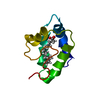
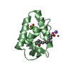
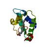
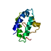
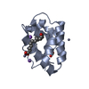
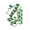
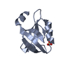
| Title | Non-specific lipid transfer protein 1 from barley in complex with L-alfa-lysophosphatidylcholine, Laudoyl | ||||||
 Components Components | nonspecific lipid-transfer protein 1 | ||||||
 Keywords Keywords | LIPID BINDING PROTEIN / lipid transfer | ||||||
| Function / homology |  Function and homology information Function and homology information | ||||||
| Biological species |  | ||||||
| Method |  X-RAY DIFFRACTION / MOLECULAR REPLACEMENT / Resolution: 1.71 Å X-RAY DIFFRACTION / MOLECULAR REPLACEMENT / Resolution: 1.71 Å | ||||||
 Authors Authors | Henriksen, A. | ||||||
 Citation Citation | Journal: To be Published Title: Binding of L-alfa-lysophosphatidylcholine, lauroyl in the hydrophobic channel of barley lipid transfer protein Authors: Henriksen, A. | ||||||
| History |
|
- Structure visualization
| Structure viewer | Molecule: MolmilJmol/JSmol |
|---|
- Downloads & links
-Download
| PDBx/mmCIF format | 1mid.cif.gz | 32.7 KB | Display | PDBx/mmCIF format |
|---|---|---|---|---|
| PDB format | pdb1mid.ent.gz | 20.8 KB | Display | PDB format |
| PDBx/mmJSON format | 1mid.json.gz | Tree view | PDBx/mmJSON format | |
| Others |  Other downloads Other downloads |
-Validation report
| Arichive directory | https://data.pdbj.org/pub/pdb/validation_reports/mi/1midftp://data.pdbj.org/pub/pdb/validation_reports/mi/1mid | HTTPS FTP |
|---|
-Related structure data
| Related structure data |  1mzlS S: Starting model for refinement |
|---|---|
| Similar structure data |


-Links
 PDBj
PDBj
- Assembly
| Deposited unit | 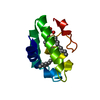
| ||||||||
|---|---|---|---|---|---|---|---|---|---|
| 1 |
| ||||||||
| Unit cell |
|
-Components
| #1: Protein | Mass: 9704.965 Da / Num. of mol.: 1 / Source method: isolated from a natural source / Source: (natural) | ||||
|---|---|---|---|---|---|
| #2: Chemical |   Mass: 440.532 Da / Num. of mol.: 3 / Source method: obtained synthetically / Formula: C20H43NO7P Mass: 440.532 Da / Num. of mol.: 3 / Source method: obtained synthetically / Formula: C20H43NO7P#3: Water | ChemComp-HOH / |  Mass: 18.015 Da / Num. of mol.: 87 / Source method: isolated from a natural source / Formula: H2O Mass: 18.015 Da / Num. of mol.: 87 / Source method: isolated from a natural source / Formula: H2OHas protein modification | Y | |
-Experimental details
-Experiment
| Experiment | Method: X-RAY DIFFRACTION / Number of used crystals: 1 |
|---|
- Sample preparation
| Crystal | Density Matthews: 1.79 Å3/Da / Density % sol: 31.44 % |
|---|---|
| Crystal grow | Temperature: 298 K / Method: vapor diffusion, hanging drop / pH: 8.5 Details: 1.1M sodium citrate, 0.1M Tris, pH 8.5, VAPOR DIFFUSION, HANGING DROP, temperature 298K |
-Data collection
| Diffraction | Mean temperature: 100 K |
|---|---|
| Diffraction source | Source: ROTATING ANODE / Type: RIGAKU RU300 / Wavelength: 1.5418 Å |
| Detector | Type: RIGAKU RAXIS IV / Detector: IMAGE PLATE / Date: Nov 2, 2000 / Details: mirrors |
| Radiation | Monochromator: Osmic mirrors / Protocol: SINGLE WAVELENGTH / Monochromatic (M) / Laue (L): M / Scattering type: x-ray |
| Radiation wavelength | Wavelength: 1.5418 Å / Relative weight: 1 |
| Reflection | Resolution: 1.71→21.93 Å / Num. all: 7578 / Num. obs: 7578 / % possible obs: 96 % / Observed criterion σ(F): 0 / Redundancy: 3.4 % / Biso Wilson estimate: 18.17 Å2 / Rmerge(I) obs: 0.056 / Net I/σ(I): 10.8 |
| Reflection shell | Resolution: 1.71→1.76 Å / Redundancy: 2.3 % / Rmerge(I) obs: 0.233 / Mean I/σ(I) obs: 4.4 / Num. unique all: 693 / % possible all: 93.6 |
- Processing
| Software |
| ||||||||||||||||||||||||||||||||||||
|---|---|---|---|---|---|---|---|---|---|---|---|---|---|---|---|---|---|---|---|---|---|---|---|---|---|---|---|---|---|---|---|---|---|---|---|---|---|
| Refinement | Method to determine structure: MOLECULAR REPLACEMENT Starting model: PDB ENTRY 1MZL Resolution: 1.71→21.93 Å / Rfactor Rfree error: 0.008 / Data cutoff high absF: 559390.06 / Data cutoff low absF: 0 / Isotropic thermal model: RESTRAINED / Cross valid method: THROUGHOUT / σ(F): 0 / Stereochemistry target values: Engh & Huber Details: The side chains of ARG 56, PRO 83 AND ASP 84 are disordered and have not been included in the model. Some atoms in the LPL ligand had no significant electron density and have been omitted from the model
| ||||||||||||||||||||||||||||||||||||
| Solvent computation | Solvent model: FLAT MODEL / Bsol: 40.2627 Å2 / ksol: 0.382403 e/Å3 | ||||||||||||||||||||||||||||||||||||
| Displacement parameters | Biso mean: 20.6 Å2
| ||||||||||||||||||||||||||||||||||||
| Refine analyze |
| ||||||||||||||||||||||||||||||||||||
| Refinement step | Cycle: LAST / Resolution: 1.71→21.93 Å
| ||||||||||||||||||||||||||||||||||||
| Refine LS restraints |
| ||||||||||||||||||||||||||||||||||||
| LS refinement shell | Resolution: 1.7→1.81 Å / Rfactor Rfree error: 0.027 / Total num. of bins used: 6
| ||||||||||||||||||||||||||||||||||||
| Xplor file |
|
