 Movie
Movie Controller

[English] 日本語
 Yorodumi
- PDB-1mfq: Crystal Structure Analysis of a Ternary S-Domain Complex of Human... -
+ Open data

- Basic information
| Entry | Database: PDB / ID: 1mfq | ||||||
|---|---|---|---|---|---|---|---|
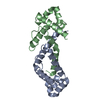
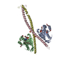
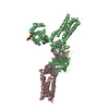
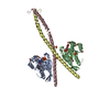
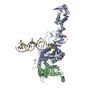
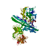
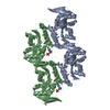
| Title | Crystal Structure Analysis of a Ternary S-Domain Complex of Human Signal Recognition Particle | ||||||
 Components Components |
| ||||||
 Keywords Keywords | SIGNALING PROTEIN/RNA / RNA-protein complex / A-minor motif / 3-helix junction / SIGNALING PROTEIN-RNA COMPLEX | ||||||
| Function / homology |  Function and homology information Function and homology informationSRP-dependent cotranslational protein targeting to membrane, signal sequence recognition / endoplasmic reticulum signal sequence receptor activity / signal recognition particle, endoplasmic reticulum targeting / signal recognition particle / cotranslational protein targeting to membrane / granulocyte differentiation / signal-recognition-particle GTPase / protein targeting to ER / SRP-dependent cotranslational protein targeting to membrane, translocation / 7S RNA binding ...SRP-dependent cotranslational protein targeting to membrane, signal sequence recognition / endoplasmic reticulum signal sequence receptor activity / signal recognition particle, endoplasmic reticulum targeting / signal recognition particle / cotranslational protein targeting to membrane / granulocyte differentiation / signal-recognition-particle GTPase / protein targeting to ER / SRP-dependent cotranslational protein targeting to membrane, translocation / 7S RNA binding / SRP-dependent cotranslational protein targeting to membrane / exocrine pancreas development / SRP-dependent cotranslational protein targeting to membrane / ribonucleoprotein complex binding / neutrophil chemotaxis / GDP binding / nuclear speck / nuclear body / GTPase activity / GTP binding / nucleolus / endoplasmic reticulum / RNA binding / nucleus / cytoplasm / cytosol Similarity search - Function | ||||||
| Biological species |  Homo sapiens (human) Homo sapiens (human) | ||||||
| Method |  X-RAY DIFFRACTION / SYNCHROTRON / SIRAS / Resolution: 3.1 Å X-RAY DIFFRACTION / SYNCHROTRON / SIRAS / Resolution: 3.1 Å | ||||||
 Authors Authors | Kuglstatter, A. / Oubridge, C. / Nagai, K. | ||||||
 Citation Citation | Journal: Nat.Struct.Biol. / Year: 2002 Title: Induced structural changes of 7SL RNA during the assembly of human signal recognition particle Authors: Kuglstatter, A. / Oubridge, C. / Nagai, K. | ||||||
| History |
|
- Structure visualization
| Structure viewer | Molecule: MolmilJmol/JSmol |
|---|
- Downloads & links
-Download
| PDBx/mmCIF format | 1mfq.cif.gz | 131 KB | Display | PDBx/mmCIF format |
|---|---|---|---|---|
| PDB format | pdb1mfq.ent.gz | 96.3 KB | Display | PDB format |
| PDBx/mmJSON format | 1mfq.json.gz | Tree view | PDBx/mmJSON format | |
| Others |  Other downloads Other downloads |
-Validation report
| Arichive directory | https://data.pdbj.org/pub/pdb/validation_reports/mf/1mfqftp://data.pdbj.org/pub/pdb/validation_reports/mf/1mfq | HTTPS FTP |
|---|
-Related structure data


-Links
 PDBj
PDBj


- Assembly
| Deposited unit | 
| ||||||||
|---|---|---|---|---|---|---|---|---|---|
| 1 |
| ||||||||
| Unit cell |
|
-Components
-RNA chain , 1 types, 1 molecules A
| #1: RNA chain | Mass: 41566.715 Da / Num. of mol.: 1 / Fragment: S-domain Source method: isolated from a genetically manipulated source Source: (gene. exp.) Homo sapiens (human) / Description: in vitro transcription with T7 RNA polymerase / Plasmid: pUC18 |
|---|
-Signal recognition particle ... , 2 types, 2 molecules BC
| #2: Protein | Mass: 12561.688 Da / Num. of mol.: 1 Source method: isolated from a genetically manipulated source Source: (gene. exp.) Homo sapiens (human) / Gene: SRP19 / Plasmid: pET23d / Production host:  |
|---|---|
| #3: Protein | Mass: 14733.043 Da / Num. of mol.: 1 / Fragment: M-domain Source method: isolated from a genetically manipulated source Source: (gene. exp.) Homo sapiens (human) / Gene: SRP54 / Plasmid: pRK172 / Species (production host): Escherichia coli / Production host: |
-Non-polymers , 3 types, 26 molecules 


| #4: Chemical | ChemComp-MG /  Mass: 24.305 Da / Num. of mol.: 5 / Source method: obtained synthetically / Formula: Mg Mass: 24.305 Da / Num. of mol.: 5 / Source method: obtained synthetically / Formula: Mg#5: Chemical |  Mass: 35.453 Da / Num. of mol.: 2 / Source method: obtained synthetically / Formula: Cl Mass: 35.453 Da / Num. of mol.: 2 / Source method: obtained synthetically / Formula: Cl#6: Water | ChemComp-HOH / | Mass: 18.015 Da / Num. of mol.: 19 / Source method: isolated from a natural source / Formula: H2O |
|---|
-Experimental details
-Experiment
| Experiment | Method: X-RAY DIFFRACTION / Number of used crystals: 1 |
|---|
- Sample preparation
| Crystal | Density Matthews: 3.68 Å3/Da / Density % sol: 66.57 % | ||||||||||||||||||||||||||||||||||||||||||||||||||||||||||||||||||||||
|---|---|---|---|---|---|---|---|---|---|---|---|---|---|---|---|---|---|---|---|---|---|---|---|---|---|---|---|---|---|---|---|---|---|---|---|---|---|---|---|---|---|---|---|---|---|---|---|---|---|---|---|---|---|---|---|---|---|---|---|---|---|---|---|---|---|---|---|---|---|---|---|
| Crystal grow | Temperature: 300.5 K / Method: vapor diffusion, hanging drop / pH: 6.5 Details: 14% (w/v) PEG8000, 100mM Na-cacodylate, 400mM lithium sulfate, 80mM magnesium chloride, pH 6.5, VAPOR DIFFUSION, HANGING DROP, temperature 300.5K | ||||||||||||||||||||||||||||||||||||||||||||||||||||||||||||||||||||||
| Components of the solutions |
| ||||||||||||||||||||||||||||||||||||||||||||||||||||||||||||||||||||||
| Crystal grow | *PLUS pH: 7.4 | ||||||||||||||||||||||||||||||||||||||||||||||||||||||||||||||||||||||
| Components of the solutions | *PLUS
|
-Data collection
| Diffraction | Mean temperature: 100 K |
|---|---|
| Diffraction source | Source: SYNCHROTRON / Site: ESRF  / Beamline: ID14-4 / Wavelength: 0.9393 Å / Beamline: ID14-4 / Wavelength: 0.9393 Å |
| Detector | Type: ADSC QUANTUM 4 / Detector: CCD / Date: Feb 19, 2002 |
| Radiation | Protocol: SINGLE WAVELENGTH / Monochromatic (M) / Laue (L): M / Scattering type: x-ray |
| Radiation wavelength | Wavelength: 0.9393 Å / Relative weight: 1 |
| Reflection | Resolution: 3.1→50 Å / Num. obs: 19354 / % possible obs: 99.7 % / Observed criterion σ(I): -3 / Redundancy: 16.6 % / Rsym value: 0.117 / Net I/σ(I): 24.3 |
| Reflection shell | Resolution: 3.1→3.21 Å / Redundancy: 15.9 % / Mean I/σ(I) obs: 6.2 / Num. unique all: 1886 / Rsym value: 0.602 / % possible all: 100 |
| Reflection | *PLUS Rmerge(I) obs: 0.117 |
| Reflection shell | *PLUS % possible obs: 100 % / Rmerge(I) obs: 0.602 |
- Processing
| Software |
| ||||||||||||||||||||||||||||||||||||
|---|---|---|---|---|---|---|---|---|---|---|---|---|---|---|---|---|---|---|---|---|---|---|---|---|---|---|---|---|---|---|---|---|---|---|---|---|---|
| Refinement | Method to determine structure: SIRAS Starting model: PDB entries 1L9A (RNA), 1JID (SRP19), 1QB2 (SRP54 M-domain) Resolution: 3.1→49.63 Å / Rfactor Rfree error: 0.008 / Isotropic thermal model: RESTRAINED / Cross valid method: THROUGHOUT / σ(F): 0 / Stereochemistry target values: Engh & Huber
| ||||||||||||||||||||||||||||||||||||
| Solvent computation | Solvent model: FLAT MODEL / Bsol: 57.551 Å2 / ksol: 0.264604 e/Å3 | ||||||||||||||||||||||||||||||||||||
| Displacement parameters | Biso mean: 97.6 Å2
| ||||||||||||||||||||||||||||||||||||
| Refine analyze | Luzzati coordinate error free: 0.43 Å / Luzzati sigma a free: 0.45 Å | ||||||||||||||||||||||||||||||||||||
| Refinement step | Cycle: LAST / Resolution: 3.1→49.63 Å
| ||||||||||||||||||||||||||||||||||||
| Refine LS restraints |
| ||||||||||||||||||||||||||||||||||||
| LS refinement shell | Resolution: 3.1→3.29 Å / Rfactor Rfree error: 0.03 / Total num. of bins used: 6
| ||||||||||||||||||||||||||||||||||||
| Xplor file |
| ||||||||||||||||||||||||||||||||||||
| Refinement | *PLUS % reflection Rfree: 5 % / Rfactor Rfree: 0.267 / Rfactor Rwork: 0.229 | ||||||||||||||||||||||||||||||||||||
| Solvent computation | *PLUS | ||||||||||||||||||||||||||||||||||||
| Displacement parameters | *PLUS | ||||||||||||||||||||||||||||||||||||
| Refine LS restraints | *PLUS
| ||||||||||||||||||||||||||||||||||||
| LS refinement shell | *PLUS Rfactor Rfree: 0.383 / Rfactor Rwork: 0.298 |
