 Movie
Movie Controller


+ Open data

- Basic information
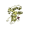
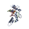
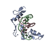
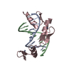
| Entry | Database: PDB / ID: 1mey | ||||||
|---|---|---|---|---|---|---|---|
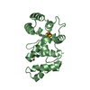
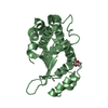
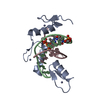
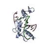
| Title | CRYSTAL STRUCTURE OF A DESIGNED ZINC FINGER PROTEIN BOUND TO DNA | ||||||
 Components Components |
| ||||||
 Keywords Keywords | TRANSFERASE/DNA / ZINC FINGER / PROTEIN-DNA INTERACTION / PROTEIN DESIGN / COMPLEX (ZINC FINGER-DNA) / TRANSFERASE-DNA COMPLEX | ||||||
| Function / homology | Classic Zinc Finger / Double Stranded RNA Binding Domain / 2-Layer Sandwich / Alpha Beta / DNA / DNA (> 10) Function and homology information Function and homology information | ||||||
| Method |  X-RAY DIFFRACTION / Resolution: 2.2 Å X-RAY DIFFRACTION / Resolution: 2.2 Å | ||||||
 Authors Authors | Kim, C.A. / Berg, J.M. | ||||||
 Citation Citation | Journal: Nat.Struct.Biol. / Year: 1996 Title: A 2.2 A Resolution Crystal Structure of a Designed Zinc Finger Protein Bound to DNA Authors: Kim, C.A. / Berg, J.M. #1: Journal: J.Mol.Biol. / Year: 1995Title: Serine at Position 2 in the DNA Recognition Helix of a Cys2-His2 Zinc Finger Peptide is not, in General, Responsible for Base Recognition Authors: Kim, C.A. / Berg, J.M. #2: Journal: Science / Year: 1993Title: Crystal Structure of a Five-Finger GLI-DNA Complex: New Perspectives on Zinc Fingers Authors: Pavletich, N.P. / Pabo, C.O. #3: Journal: Nature / Year: 1993Title: The Crystal Structure of a Two Zinc-Finger Peptide Reveals an Extension to the Rules for Zinc-Finger/DNA Recognition Authors: Fairall, L. / Schwabe, J.W. / Chapman, L. / Finch, J.T. / Rhodes, D. #4: Journal: Science / Year: 1991Title: Zinc Finger-DNA Recognition: Crystal Structure of a Zif268-DNA Complex at 2.1 A Authors: Pavletich, N.P. / Pabo, C.O. | ||||||
| History |
|
- Structure visualization
| Structure viewer | Molecule: MolmilJmol/JSmol |
|---|
- Downloads & links
-Download
| PDBx/mmCIF format | 1mey.cif.gz | 86.1 KB | Display | PDBx/mmCIF format |
|---|---|---|---|---|
| PDB format | pdb1mey.ent.gz | 62.4 KB | Display | PDB format |
| PDBx/mmJSON format | 1mey.json.gz | Tree view | PDBx/mmJSON format | |
| Others |  Other downloads Other downloads |
-Validation report
| Arichive directory | https://data.pdbj.org/pub/pdb/validation_reports/me/1meyftp://data.pdbj.org/pub/pdb/validation_reports/me/1mey | HTTPS FTP |
|---|
-Related structure data
| Similar structure data |
|---|


-Links
 PDBj
PDBj


- Assembly
| Deposited unit | 
| ||||||||||
|---|---|---|---|---|---|---|---|---|---|---|---|
| 1 | 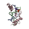
| ||||||||||
| 2 | 
| ||||||||||
| 3 | 
| ||||||||||
| Unit cell |
|
-Components
-DNA chain , 2 types, 4 molecules ADBE
| #1: DNA chain | Mass: 4024.649 Da / Num. of mol.: 2 / Source method: obtained synthetically #2: DNA chain | Mass: 4043.456 Da / Num. of mol.: 2 / Source method: obtained synthetically |
|---|
-Protein , 1 types, 3 molecules CFG
| #3: Protein | Mass: 10147.411 Da / Num. of mol.: 3 Source method: isolated from a genetically manipulated source Species (production host): Escherichia coli / Production host:  |
|---|
-Non-polymers , 3 types, 141 molecules 


| #4: Chemical | ChemComp-ZN /  Mass: 65.409 Da / Num. of mol.: 8 / Source method: obtained synthetically / Formula: Zn Mass: 65.409 Da / Num. of mol.: 8 / Source method: obtained synthetically / Formula: Zn#5: Chemical | ChemComp-CL / |  Mass: 35.453 Da / Num. of mol.: 1 / Source method: obtained synthetically / Formula: Cl Mass: 35.453 Da / Num. of mol.: 1 / Source method: obtained synthetically / Formula: Cl#6: Water | ChemComp-HOH / | Mass: 18.015 Da / Num. of mol.: 132 / Source method: isolated from a natural source / Formula: H2O |
|---|
-Details
| Compound details | THE ENTRY CONTAINS TWO COMPLEXES AND ONE UNPAIRED PROTEIN MOLECULE WHICH MAKE UP THE ASYMMETRIC ...THE ENTRY CONTAINS TWO COMPLEXES AND ONE UNPAIRED PROTEIN MOLECULE WHICH MAKE UP THE ASYMMETRIC |
|---|
-Experimental details
-Experiment
| Experiment | Method: X-RAY DIFFRACTION |
|---|
- Sample preparation
| Crystal | Density Matthews: 2.52 Å3/Da / Density % sol: 51.18 % | ||||||||||||||||||||||||||||||||||||
|---|---|---|---|---|---|---|---|---|---|---|---|---|---|---|---|---|---|---|---|---|---|---|---|---|---|---|---|---|---|---|---|---|---|---|---|---|---|
| Crystal grow | Temperature: 293 K / Method: vapor diffusion, hanging drop / pH: 7.8 Details: pH 7.80, VAPOR DIFFUSION, HANGING DROP, temperature 293.00K | ||||||||||||||||||||||||||||||||||||
| Components of the solutions |
| ||||||||||||||||||||||||||||||||||||
| Crystal grow | *PLUS Temperature: 20 ℃ / PH range low: 7.8 / PH range high: 7.6 | ||||||||||||||||||||||||||||||||||||
| Components of the solutions | *PLUS
|
-Data collection
| Diffraction source | Source: ROTATING ANODE |
|---|---|
| Detector | Type: RIGAKU RAXIS IIC / Detector: IMAGE PLATE |
| Radiation | Protocol: SINGLE WAVELENGTH / Monochromatic (M) / Laue (L): M / Scattering type: x-ray |
| Radiation wavelength | Relative weight: 1 |
| Reflection | Num. obs: 23392 / % possible obs: 84 % / Redundancy: 6 % / Rmerge(I) obs: 0.088 |
| Reflection | *PLUS Highest resolution: 2.2 Å / Num. measured all: 146523 |
- Processing
| Software |
| ||||||||||||||||||||||||||||||||||||||||||||||||||||||||||||
|---|---|---|---|---|---|---|---|---|---|---|---|---|---|---|---|---|---|---|---|---|---|---|---|---|---|---|---|---|---|---|---|---|---|---|---|---|---|---|---|---|---|---|---|---|---|---|---|---|---|---|---|---|---|---|---|---|---|---|---|---|---|
| Refinement | Resolution: 2.2→6 Å / σ(F): 3
| ||||||||||||||||||||||||||||||||||||||||||||||||||||||||||||
| Displacement parameters | Biso mean: 32.1 Å2 | ||||||||||||||||||||||||||||||||||||||||||||||||||||||||||||
| Refinement step | Cycle: LAST / Resolution: 2.2→6 Å
| ||||||||||||||||||||||||||||||||||||||||||||||||||||||||||||
| Refine LS restraints |
| ||||||||||||||||||||||||||||||||||||||||||||||||||||||||||||
| LS refinement shell | Resolution: 2.2→2.22 Å
| ||||||||||||||||||||||||||||||||||||||||||||||||||||||||||||
| Xplor file |
| ||||||||||||||||||||||||||||||||||||||||||||||||||||||||||||
| Software | *PLUS Name: X-PLOR / Classification: refinement | ||||||||||||||||||||||||||||||||||||||||||||||||||||||||||||
| Refinement | *PLUS Highest resolution: 2.2 Å / Lowest resolution: 6 Å / Num. reflection all: 21015 / σ(F): 3 / Rfactor all: 0.235 | ||||||||||||||||||||||||||||||||||||||||||||||||||||||||||||
| Solvent computation | *PLUS | ||||||||||||||||||||||||||||||||||||||||||||||||||||||||||||
| Displacement parameters | *PLUS | ||||||||||||||||||||||||||||||||||||||||||||||||||||||||||||
| Refine LS restraints | *PLUS
|
