 Movie
Movie Controller


+ Open data

- Basic information
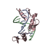
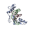
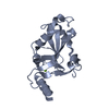
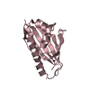
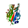
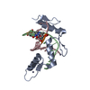
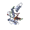
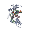
| Entry | Database: PDB / ID: 1a1j | ||||||
|---|---|---|---|---|---|---|---|
| Title | RADR (ZIF268 VARIANT) ZINC FINGER-DNA COMPLEX (GCGT SITE) | ||||||
 Components Components |
| ||||||
 Keywords Keywords | TRANSCRIPTION/DNA / ZINC FINGER-DNA COMPLEX / ZINC FINGER / DNA-BINDING PROTEIN / TRANSCRIPTION-DNA COMPLEX | ||||||
| Function / homology |  Function and homology information Function and homology informationregulation of protein sumoylation / glomerular mesangial cell proliferation / positive regulation of glomerular metanephric mesangial cell proliferation / cellular response to interleukin-8 / positive regulation of post-translational protein modification / regulation of progesterone biosynthetic process / cellular response to heparin / cellular response to mycophenolic acid / circadian temperature homeostasis / positive regulation of hormone biosynthetic process ...regulation of protein sumoylation / glomerular mesangial cell proliferation / positive regulation of glomerular metanephric mesangial cell proliferation / cellular response to interleukin-8 / positive regulation of post-translational protein modification / regulation of progesterone biosynthetic process / cellular response to heparin / cellular response to mycophenolic acid / circadian temperature homeostasis / positive regulation of hormone biosynthetic process / double-stranded methylated DNA binding / hemi-methylated DNA-binding / positive regulation of gene expression via chromosomal CpG island demethylation / interleukin-1-mediated signaling pathway / positive regulation of smooth muscle cell migration / histone acetyltransferase binding / locomotor rhythm / T cell differentiation / skeletal muscle cell differentiation / BMP signaling pathway / response to glucose / estrous cycle / RNA polymerase II core promoter sequence-specific DNA binding / long-term memory / positive regulation of chemokine production / positive regulation of smooth muscle cell proliferation / regulation of neuron apoptotic process / response to ischemia / positive regulation of interleukin-1 beta production / RNA polymerase II transcription regulatory region sequence-specific DNA binding / circadian regulation of gene expression / promoter-specific chromatin binding / negative regulation of canonical Wnt signaling pathway / cellular response to gamma radiation / response to insulin / positive regulation of miRNA transcription / regulation of long-term neuronal synaptic plasticity / sequence-specific double-stranded DNA binding / positive regulation of neuron apoptotic process / double-stranded DNA binding / DNA-binding transcription activator activity, RNA polymerase II-specific / regulation of apoptotic process / sequence-specific DNA binding / DNA-binding transcription factor activity, RNA polymerase II-specific / learning or memory / response to hypoxia / transcription cis-regulatory region binding / RNA polymerase II cis-regulatory region sequence-specific DNA binding / DNA-binding transcription factor activity / positive regulation of gene expression / regulation of transcription by RNA polymerase II / regulation of DNA-templated transcription / positive regulation of DNA-templated transcription / chromatin / enzyme binding / negative regulation of transcription by RNA polymerase II / positive regulation of transcription by RNA polymerase II / DNA binding / zinc ion binding / nucleoplasm / nucleus / cytoplasm Similarity search - Function | ||||||
| Biological species |  | ||||||
| Method |  X-RAY DIFFRACTION / ISOMORPHOUS MOLECULAR REPLACEMENT / Resolution: 2 Å X-RAY DIFFRACTION / ISOMORPHOUS MOLECULAR REPLACEMENT / Resolution: 2 Å | ||||||
 Authors Authors | Elrod-Erickson, M. / Benson, T.E. / Pabo, C.O. | ||||||
 Citation Citation | Journal: Structure / Year: 1998 Title: High-resolution structures of variant Zif268-DNA complexes: implications for understanding zinc finger-DNA recognition. Authors: Elrod-Erickson, M. / Benson, T.E. / Pabo, C.O. #1: Journal: Structure / Year: 1996Title: Zif268 Protein-DNA Complex Refined at 1.6 A: A Model System for Understanding Zinc Finger-DNA Interactions Authors: Elrod-Erickson, M. / Rould, M.A. / Nekludova, L. / Pabo, C.O. #2: Journal: Science / Year: 1994Title: Zinc Finger Phage: Affinity Selection of Fingers with New DNA-Binding Specificities Authors: Rebar, E.J. / Pabo, C.O. #3: Journal: Science / Year: 1991Title: Zinc Finger-DNA Recognition: Crystal Structure of a Zif268-DNA Complex at 2.1 A Authors: Pavletich, N.P. / Pabo, C.O. | ||||||
| History |
|
- Structure visualization
| Structure viewer | Molecule: MolmilJmol/JSmol |
|---|
- Downloads & links
-Download
| PDBx/mmCIF format | 1a1j.cif.gz | 48.6 KB | Display | PDBx/mmCIF format |
|---|---|---|---|---|
| PDB format | pdb1a1j.ent.gz | 31.3 KB | Display | PDB format |
| PDBx/mmJSON format | 1a1j.json.gz | Tree view | PDBx/mmJSON format | |
| Others |  Other downloads Other downloads |
-Validation report
| Arichive directory | https://data.pdbj.org/pub/pdb/validation_reports/a1/1a1jftp://data.pdbj.org/pub/pdb/validation_reports/a1/1a1j | HTTPS FTP |
|---|
-Related structure data
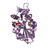
| Related structure data |  1a1fC  1a1gC  1a1hC  1a1iC  1a1kC  1a1lC  1aayS S: Starting model for refinement C: citing same article ( |
|---|---|
| Similar structure data |

-Links
 PDBj
PDBj


- Assembly
| Deposited unit | 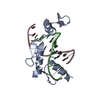
| ||||||||||
|---|---|---|---|---|---|---|---|---|---|---|---|
| 1 |
| ||||||||||
| Unit cell |
|
-Components
| #1: DNA chain | Mass: 3430.233 Da / Num. of mol.: 1 / Source method: obtained synthetically | ||
|---|---|---|---|
| #2: DNA chain | Mass: 3279.151 Da / Num. of mol.: 1 / Source method: obtained synthetically | ||
| #3: Protein | Mass: 10795.463 Da / Num. of mol.: 1 / Fragment: ZINC FINGER Source method: isolated from a genetically manipulated source Source: (gene. exp.)  | ||
| #4: Chemical |   Mass: 65.409 Da / Num. of mol.: 3 / Source method: obtained synthetically / Formula: Zn Mass: 65.409 Da / Num. of mol.: 3 / Source method: obtained synthetically / Formula: Zn#5: Water | ChemComp-HOH / |  Mass: 18.015 Da / Num. of mol.: 153 / Source method: isolated from a natural source / Formula: H2O Mass: 18.015 Da / Num. of mol.: 153 / Source method: isolated from a natural source / Formula: H2O |
-Experimental details
-Experiment
| Experiment | Method: X-RAY DIFFRACTION / Number of used crystals: 1 |
|---|
- Sample preparation
| Crystal | Density Matthews: 2.26 Å3/Da / Density % sol: 45.48 % | ||||||||||||||||||||||||||||||||||||||||||||||||
|---|---|---|---|---|---|---|---|---|---|---|---|---|---|---|---|---|---|---|---|---|---|---|---|---|---|---|---|---|---|---|---|---|---|---|---|---|---|---|---|---|---|---|---|---|---|---|---|---|---|
| Crystal grow | Method: vapor diffusion, hanging drop / pH: 6.2 Details: 35% PEG 3350, 200MM NACL, 25 MM MES PH 6.2, VAPOR DIFFUSION, HANGING DROP | ||||||||||||||||||||||||||||||||||||||||||||||||
| Components of the solutions |
| ||||||||||||||||||||||||||||||||||||||||||||||||
| Crystal grow | *PLUS | ||||||||||||||||||||||||||||||||||||||||||||||||
| Components of the solutions | *PLUS
|
-Data collection
| Diffraction | Mean temperature: 130 K |
|---|---|
| Diffraction source | Source: ROTATING ANODE / Type: RIGAKU RU200 |
| Detector | Type: RIGAKU RAXIS IIC / Detector: IMAGE PLATE / Date: Dec 15, 1995 / Details: YALE MIRRORS |
| Radiation | Monochromator: YALE MIRRORS / Monochromatic (M) / Laue (L): M / Scattering type: x-ray |
| Radiation wavelength | Relative weight: 1 |
| Reflection | Resolution: 2→20 Å / Num. obs: 9959 / % possible obs: 89.6 % / Observed criterion σ(I): -2 / Redundancy: 3 % / Rsym value: 0.034 / Net I/σ(I): 39.5 |
| Reflection shell | Resolution: 2→2.07 Å / Redundancy: 1.8 % / Mean I/σ(I) obs: 19.4 / Rsym value: 0.054 / % possible all: 82.9 |
| Reflection | *PLUS Highest resolution: 2 Å / Lowest resolution: 20 Å / % possible obs: 89.6 % / Redundancy: 3 % / Num. measured all: 29842 / Rmerge(I) obs: 0.034 |
| Reflection shell | *PLUS Highest resolution: 2 Å / Lowest resolution: 2.07 Å / % possible obs: 82.9 % / Rmerge(I) obs: 0.054 |
- Processing
| Software |
| ||||||||||||||||||||||||||||||||||||||||||||||||||||||||||||||||||||||||||||||||
|---|---|---|---|---|---|---|---|---|---|---|---|---|---|---|---|---|---|---|---|---|---|---|---|---|---|---|---|---|---|---|---|---|---|---|---|---|---|---|---|---|---|---|---|---|---|---|---|---|---|---|---|---|---|---|---|---|---|---|---|---|---|---|---|---|---|---|---|---|---|---|---|---|---|---|---|---|---|---|---|---|---|
| Refinement | Method to determine structure: ISOMORPHOUS MOLECULAR REPLACEMENT Starting model: PDB ENTRY 1AAY, WITHOUT WATERS AND WITHOUT SIDE CHAINS FOR RESIDUES 18 - 24 Resolution: 2→20 Å / Rfactor Rfree error: 0.008 / Data cutoff high absF: 100000 / Data cutoff low absF: 0 / Isotropic thermal model: RESTRAINED INDIVIDUAL / Cross valid method: THROUGHOUT / σ(F): 0
| ||||||||||||||||||||||||||||||||||||||||||||||||||||||||||||||||||||||||||||||||
| Displacement parameters | Biso mean: 32.2 Å2
| ||||||||||||||||||||||||||||||||||||||||||||||||||||||||||||||||||||||||||||||||
| Refinement step | Cycle: LAST / Resolution: 2→20 Å
| ||||||||||||||||||||||||||||||||||||||||||||||||||||||||||||||||||||||||||||||||
| Refine LS restraints |
| ||||||||||||||||||||||||||||||||||||||||||||||||||||||||||||||||||||||||||||||||
| LS refinement shell | Resolution: 2→2.09 Å / Rfactor Rfree error: 0.03 / Total num. of bins used: 8
| ||||||||||||||||||||||||||||||||||||||||||||||||||||||||||||||||||||||||||||||||
| Xplor file |
| ||||||||||||||||||||||||||||||||||||||||||||||||||||||||||||||||||||||||||||||||
| Software | *PLUS Name: X-PLOR / Version: 3.8 / Classification: refinement | ||||||||||||||||||||||||||||||||||||||||||||||||||||||||||||||||||||||||||||||||
| Refinement | *PLUS Highest resolution: 2 Å / Lowest resolution: 20 Å / σ(F): 0 / % reflection Rfree: 10.9 % | ||||||||||||||||||||||||||||||||||||||||||||||||||||||||||||||||||||||||||||||||
| Solvent computation | *PLUS | ||||||||||||||||||||||||||||||||||||||||||||||||||||||||||||||||||||||||||||||||
| Displacement parameters | *PLUS | ||||||||||||||||||||||||||||||||||||||||||||||||||||||||||||||||||||||||||||||||
| Refine LS restraints | *PLUS
| ||||||||||||||||||||||||||||||||||||||||||||||||||||||||||||||||||||||||||||||||
| LS refinement shell | *PLUS Rfactor obs: 0.299 |
