 Movie
Movie Controller

[English] 日本語
 Yorodumi
- PDB-1maw: Crystal Structure of Tryptophanyl-tRNA Synthetase Complexed with ... -
+ Open data

- Basic information
| Entry | Database: PDB / ID: 1maw | ||||||
|---|---|---|---|---|---|---|---|
| Title | Crystal Structure of Tryptophanyl-tRNA Synthetase Complexed with ATP in an Open Conformation | ||||||
 Components Components | TRYPTOPHAN-TRNA LIGASE | ||||||
 Keywords Keywords | LIGASE / Amino-acyl tRNA synthetase / Rossmann fold / atp binding site | ||||||
| Function / homology |  Function and homology information Function and homology informationtryptophan-tRNA ligase / tryptophanyl-tRNA aminoacylation / tryptophan-tRNA ligase activity / ATP binding / cytosol Similarity search - Function | ||||||
| Biological species |   Geobacillus stearothermophilus (bacteria) Geobacillus stearothermophilus (bacteria) | ||||||
| Method |  X-RAY DIFFRACTION / MOLECULAR REPLACEMENT / Resolution: 3 Å X-RAY DIFFRACTION / MOLECULAR REPLACEMENT / Resolution: 3 Å | ||||||
 Authors Authors | Retailleau, P. / Huang, X. / Yin, Y. / Hu, M. / Weinreb, V. / Vachette, P. / Vonrhein, C. / Bricogne, G. / Roversi, P. / Ilyin, V. / Carter Jr., C.W. | ||||||
 Citation Citation | Journal: J.Mol.Biol. / Year: 2003 Title: Interconversion of ATP binding and conformational free energies by tryptophanyl-tRNA synthetase: structures of ATP bound to open and closed, pre-transition-state conformations. Authors: Retailleau, P. / Huang, X. / Yin, Y. / Hu, M. / Weinreb, V. / Vachette, P. / Vonrhein, C. / Bricogne, G. / Roversi, P. / Ilyin, V. / Carter, C.W. | ||||||
| History |
|
- Structure visualization
| Structure viewer | Molecule: MolmilJmol/JSmol |
|---|
- Downloads & links
-Download
| PDBx/mmCIF format | 1maw.cif.gz | 380.6 KB | Display | PDBx/mmCIF format |
|---|---|---|---|---|
| PDB format | pdb1maw.ent.gz | 313.3 KB | Display | PDB format |
| PDBx/mmJSON format | 1maw.json.gz | Tree view | PDBx/mmJSON format | |
| Others |  Other downloads Other downloads |
-Validation report
| Arichive directory | https://data.pdbj.org/pub/pdb/validation_reports/ma/1mawftp://data.pdbj.org/pub/pdb/validation_reports/ma/1maw | HTTPS FTP |
|---|
-Related structure data
| Related structure data |  1m83C  1mauC  1mb2C  1d2rS S: Starting model for refinement C: citing same article ( |
|---|---|
| Similar structure data |


-Links
 PDBj
PDBj
- Assembly
| Deposited unit | 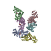
| ||||||||
|---|---|---|---|---|---|---|---|---|---|
| 1 | 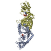
| ||||||||
| 2 | 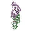
| ||||||||
| 3 | 
| ||||||||
| Unit cell |
|
-Components
| #1: Protein | Mass: 37225.672 Da / Num. of mol.: 6 Source method: isolated from a genetically manipulated source Source: (gene. exp.) Geobacillus stearothermophilus (bacteria)Production host: #2: Chemical | ChemComp-ATP /   Mass: 507.181 Da / Num. of mol.: 6 / Source method: obtained synthetically / Formula: C10H16N5O13P3 / Comment: ATP, energy-carrying molecule*YM Mass: 507.181 Da / Num. of mol.: 6 / Source method: obtained synthetically / Formula: C10H16N5O13P3 / Comment: ATP, energy-carrying molecule*YM |
|---|
-Experimental details
-Experiment
| Experiment | Method: X-RAY DIFFRACTION / Number of used crystals: 1 |
|---|
- Sample preparation
| Crystal | Density Matthews: 2.73 Å3/Da / Density % sol: 54.91 % | |||||||||||||||||||||||||||||||||||
|---|---|---|---|---|---|---|---|---|---|---|---|---|---|---|---|---|---|---|---|---|---|---|---|---|---|---|---|---|---|---|---|---|---|---|---|---|
| Crystal grow | Temperature: 310 K / Method: microdialysis / pH: 6.6 Details: potassium phosphate, mg-atp, pH 6.6, MICRODIALYSIS, temperature 310K | |||||||||||||||||||||||||||||||||||
| Crystal grow | *PLUS Temperature: 42 ℃ | |||||||||||||||||||||||||||||||||||
| Components of the solutions | *PLUS
|
-Data collection
| Diffraction | Mean temperature: 298 K |
|---|---|
| Diffraction source | Source: ROTATING ANODE / Type: RIGAKU RU200 / Wavelength: 1.5418 Å |
| Detector | Type: RIGAKU RAXIS II / Detector: IMAGE PLATE |
| Radiation | Monochromator: GRAPHITE / Protocol: SINGLE WAVELENGTH / Monochromatic (M) / Laue (L): M / Scattering type: x-ray |
| Radiation wavelength | Wavelength: 1.5418 Å / Relative weight: 1 |
| Reflection | Resolution: 2.99→15.05 Å / Num. obs: 42607 / % possible obs: 87.1 % / Biso Wilson estimate: 48.3 Å2 / Rsym value: 0.079 |
| Reflection | *PLUS Highest resolution: 3 Å / % possible obs: 87.3 % / Num. measured all: 102635 / Rmerge(I) obs: 0.079 |
| Reflection shell | *PLUS Highest resolution: 3 Å / Lowest resolution: 3.11 Å / % possible obs: 70.5 % |
- Processing
| Software |
| ||||||||||||||||||||||||||||||||||||
|---|---|---|---|---|---|---|---|---|---|---|---|---|---|---|---|---|---|---|---|---|---|---|---|---|---|---|---|---|---|---|---|---|---|---|---|---|---|
| Refinement | Method to determine structure: MOLECULAR REPLACEMENT Starting model: PDB ENTRY 1D2R Resolution: 3→15 Å / Rfactor Rfree error: 0.004 / Cross valid method: THROUGHOUT / σ(F): 0 / Stereochemistry target values: Engh & Huber
| ||||||||||||||||||||||||||||||||||||
| Displacement parameters | Biso mean: 41.1 Å2
| ||||||||||||||||||||||||||||||||||||
| Refine analyze |
| ||||||||||||||||||||||||||||||||||||
| Refinement step | Cycle: LAST / Resolution: 3→15 Å
| ||||||||||||||||||||||||||||||||||||
| Refine LS restraints |
| ||||||||||||||||||||||||||||||||||||
| LS refinement shell | Resolution: 3→3.19 Å / Rfactor Rfree error: 0.014 / Total num. of bins used: 6
| ||||||||||||||||||||||||||||||||||||
| Xplor file |
| ||||||||||||||||||||||||||||||||||||
| Refinement | *PLUS Highest resolution: 3 Å / Lowest resolution: 15 Å / Num. reflection obs: 38186 / % reflection Rfree: 10 % / Rfactor Rfree: 0.239 / Rfactor Rwork: 0.154 | ||||||||||||||||||||||||||||||||||||
| Solvent computation | *PLUS | ||||||||||||||||||||||||||||||||||||
| Displacement parameters | *PLUS | ||||||||||||||||||||||||||||||||||||
| Refine LS restraints | *PLUS
|
