 Movie
Movie Controller

[English] 日本語
 Yorodumi
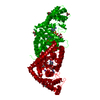
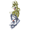
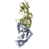
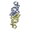
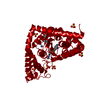
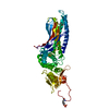
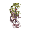
- PDB-1m83: Crystal Structure of Tryptophanyl-tRNA Synthetase Complexed with ... -
+ Open data

- Basic information
| Entry | Database: PDB / ID: 1m83 | ||||||
|---|---|---|---|---|---|---|---|
| Title | Crystal Structure of Tryptophanyl-tRNA Synthetase Complexed with ATP in a Closed, Pre-transition State Conformation | ||||||
 Components Components | Tryptophan-tRNA ligase | ||||||
 Keywords Keywords | LIGASE / Aminoacyl-tRNA Synthetase / ATP binding site / Rossmann Fold | ||||||
| Function / homology |  Function and homology information Function and homology informationtryptophan-tRNA ligase / tryptophanyl-tRNA aminoacylation / tryptophan-tRNA ligase activity / ATP binding / cytosol Similarity search - Function | ||||||
| Biological species |   Geobacillus stearothermophilus (bacteria) Geobacillus stearothermophilus (bacteria) | ||||||
| Method |  X-RAY DIFFRACTION / SIR / Resolution: 2.2 Å X-RAY DIFFRACTION / SIR / Resolution: 2.2 Å | ||||||
 Authors Authors | Retailleau, P. / Huang, X. / Yin, Y. / Hu, M. / Weinreb, V. / Vachette, P. / Vonrhein, C. / Bricogne, G. / Roversi, P. / Ilyin, V. / Carter Jr., C.W. | ||||||
 Citation Citation | Journal: J.Mol.Biol. / Year: 2003 Title: Interconversion of ATP binding and conformational free energies by tryptophanyl-tRNA synthetase: structures of ATP bound to open and closed, pre-transition-state conformations. Authors: Retailleau, P. / Huang, X. / Yin, Y. / Hu, M. / Weinreb, V. / Vachette, P. / Vonrhein, C. / Bricogne, G. / Roversi, P. / Ilyin, V. / Carter, C.W. | ||||||
| History |
|
- Structure visualization
| Structure viewer | Molecule: MolmilJmol/JSmol |
|---|
- Downloads & links
-Download
| PDBx/mmCIF format | 1m83.cif.gz | 82.4 KB | Display | PDBx/mmCIF format |
|---|---|---|---|---|
| PDB format | pdb1m83.ent.gz | 62 KB | Display | PDB format |
| PDBx/mmJSON format | 1m83.json.gz | Tree view | PDBx/mmJSON format | |
| Others |  Other downloads Other downloads |
-Validation report
| Arichive directory | https://data.pdbj.org/pub/pdb/validation_reports/m8/1m83ftp://data.pdbj.org/pub/pdb/validation_reports/m8/1m83 | HTTPS FTP |
|---|
-Related structure data


-Links
 PDBj
PDBj
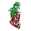
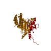
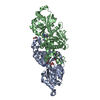
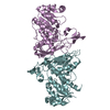
- Assembly
| Deposited unit | 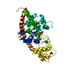
| ||||||||
|---|---|---|---|---|---|---|---|---|---|
| 1 | 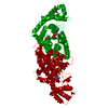
| ||||||||
| Unit cell |
|
-Components
| #1: Protein | Mass: 37225.672 Da / Num. of mol.: 1 Source method: isolated from a genetically manipulated source Source: (gene. exp.) Geobacillus stearothermophilus (bacteria)Production host: | ||
|---|---|---|---|
| #2: Chemical | ChemComp-MG /   Mass: 24.305 Da / Num. of mol.: 1 / Source method: obtained synthetically / Formula: Mg Mass: 24.305 Da / Num. of mol.: 1 / Source method: obtained synthetically / Formula: Mg | ||
| #3: Chemical | ChemComp-ATP /   Mass: 507.181 Da / Num. of mol.: 1 / Source method: obtained synthetically / Formula: C10H16N5O13P3 / Comment: ATP, energy-carrying molecule*YM Mass: 507.181 Da / Num. of mol.: 1 / Source method: obtained synthetically / Formula: C10H16N5O13P3 / Comment: ATP, energy-carrying molecule*YM | ||
| #4: Chemical |   Mass: 92.094 Da / Num. of mol.: 3 / Source method: obtained synthetically / Formula: C3H8O3 Mass: 92.094 Da / Num. of mol.: 3 / Source method: obtained synthetically / Formula: C3H8O3#5: Water | ChemComp-HOH / |  Mass: 18.015 Da / Num. of mol.: 168 / Source method: isolated from a natural source / Formula: H2O Mass: 18.015 Da / Num. of mol.: 168 / Source method: isolated from a natural source / Formula: H2O |
-Experimental details
-Experiment
| Experiment | Method: X-RAY DIFFRACTION / Number of used crystals: 2 |
|---|
- Sample preparation
| Crystal | Density Matthews: 2.64 Å3/Da / Density % sol: 47 % | ||||||||||||||||||||||||||||||||||||||||||||||||
|---|---|---|---|---|---|---|---|---|---|---|---|---|---|---|---|---|---|---|---|---|---|---|---|---|---|---|---|---|---|---|---|---|---|---|---|---|---|---|---|---|---|---|---|---|---|---|---|---|---|
| Crystal grow | Temperature: 310 K / Method: microdialysis / pH: 7.5 Details: sodium citrate, sodium atp, magnesium chloride, pH 7.5, MICRODIALYSIS, temperature 310K | ||||||||||||||||||||||||||||||||||||||||||||||||
| Crystal grow | *PLUS Temperature: 35 ℃ | ||||||||||||||||||||||||||||||||||||||||||||||||
| Components of the solutions | *PLUS
|
-Data collection
| Diffraction | Mean temperature: 100 K |
|---|---|
| Diffraction source | Source: ROTATING ANODE / Type: RIGAKU / Wavelength: 1.5418 Å |
| Detector | Type: RIGAKU RAXIS IV / Detector: IMAGE PLATE / Date: Apr 1, 1999 |
| Radiation | Monochromator: GRAPHITE / Protocol: SIR / Monochromatic (M) / Laue (L): M / Scattering type: x-ray |
| Radiation wavelength | Wavelength: 1.5418 Å / Relative weight: 1 |
| Reflection | Resolution: 2.15→45 Å / Num. all: 24329 / Num. obs: 19650 / % possible obs: 80.8 % / Observed criterion σ(I): -3 / Redundancy: 8.2 % / Biso Wilson estimate: 32.1 Å2 / Rsym value: 0.08 / Net I/σ(I): 10.2 |
| Reflection shell | Resolution: 2.15→2.23 Å / Redundancy: 1.9 % / Mean I/σ(I) obs: 2.3 / Num. unique all: 285 / Rsym value: 0.31 / % possible all: 12.1 |
| Reflection | *PLUS Lowest resolution: 45 Å / Num. obs: 24329 / % possible obs: 97.5 % / Num. measured all: 303099 / Rmerge(I) obs: 0.08 |
| Reflection shell | *PLUS % possible obs: 12.1 % / Rmerge(I) obs: 0.31 |
- Processing
| Software |
| ||||||||||||||||||||||||||||
|---|---|---|---|---|---|---|---|---|---|---|---|---|---|---|---|---|---|---|---|---|---|---|---|---|---|---|---|---|---|
| Refinement | Method to determine structure: SIR / Resolution: 2.2→20 Å / Stereochemistry target values: Engh & Huber
| ||||||||||||||||||||||||||||
| Refinement step | Cycle: LAST / Resolution: 2.2→20 Å
| ||||||||||||||||||||||||||||
| Refine LS restraints |
| ||||||||||||||||||||||||||||
| LS refinement shell |
| ||||||||||||||||||||||||||||
| Refinement | *PLUS Lowest resolution: 20 Å / % reflection Rfree: 10 % | ||||||||||||||||||||||||||||
| Solvent computation | *PLUS | ||||||||||||||||||||||||||||
| Displacement parameters | *PLUS |
