 Movie
Movie Controller


+ Open data

- Basic information
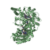
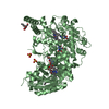
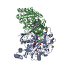
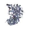
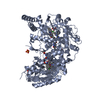
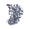
| Entry | Database: PDB / ID: 1m8d | ||||||
|---|---|---|---|---|---|---|---|
| Title | inducible nitric oxide synthase with Chlorzoxazone bound | ||||||
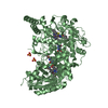
 Components Components | Inducible Nitric Oxide Synthase | ||||||
 Keywords Keywords | OXIDOREDUCTASE / inhibitor-induced conformational change | ||||||
| Function / homology |  Function and homology information Function and homology informationNitric oxide stimulates guanylate cyclase / ROS and RNS production in phagocytes / peptidyl-cysteine S-nitrosylation / G protein-coupled receptor signaling pathway coupled to cGMP nucleotide second messenger / cAMP-dependent protein kinase regulator activity / Peroxisomal protein import / prostaglandin secretion / tetrahydrobiopterin binding / : / arginine binding ...Nitric oxide stimulates guanylate cyclase / ROS and RNS production in phagocytes / peptidyl-cysteine S-nitrosylation / G protein-coupled receptor signaling pathway coupled to cGMP nucleotide second messenger / cAMP-dependent protein kinase regulator activity / Peroxisomal protein import / prostaglandin secretion / tetrahydrobiopterin binding / : / arginine binding / superoxide metabolic process / regulation of cytokine production involved in inflammatory response / Fc-gamma receptor signaling pathway involved in phagocytosis / cellular response to cytokine stimulus / regulation of insulin secretion / cortical cytoskeleton / nitric-oxide synthase binding / response to tumor necrosis factor / nitric-oxide synthase (NADPH) / : / blood vessel remodeling / nitric-oxide synthase activity / L-arginine catabolic process / nitric oxide biosynthetic process / negative regulation of blood pressure / response to hormone / positive regulation of interleukin-8 production / response to bacterium / circadian rhythm / negative regulation of protein catabolic process / Hsp90 protein binding / positive regulation of interleukin-6 production / beta-catenin binding / cellular response to type II interferon / regulation of blood pressure / cellular response to xenobiotic stimulus / NADP binding / FMN binding / flavin adenine dinucleotide binding / peroxisome / regulation of cell population proliferation / cellular response to lipopolysaccharide / actin binding / response to lipopolysaccharide / response to hypoxia / calmodulin binding / defense response to bacterium / intracellular signal transduction / cadherin binding / positive regulation of apoptotic process / inflammatory response / negative regulation of gene expression / heme binding / protein kinase binding / perinuclear region of cytoplasm / protein homodimerization activity / : / metal ion binding / identical protein binding / nucleus / plasma membrane / cytoplasm / cytosol Similarity search - Function | ||||||
| Biological species |  | ||||||
| Method |  X-RAY DIFFRACTION / SYNCHROTRON / MOLECULAR REPLACEMENT / Resolution: 2.35 Å X-RAY DIFFRACTION / SYNCHROTRON / MOLECULAR REPLACEMENT / Resolution: 2.35 Å | ||||||
 Authors Authors | Rosenfeld, R.J. / Garcin, E.D. / Panda, K. / Andersson, G. / Aberg, A. / Wallace, A.V. / Stuehr, D.J. / Tainer, J.A. / Getzoff, E.D. | ||||||
 Citation Citation | Journal: Biochemistry / Year: 2002 Title: Conformational Changes in Nitric Oxide Synthases Induced by Chlorzoxazone and Nitroindazoles: Crystallographic and Computational Analyses of Inhibitor Potency Authors: Rosenfeld, R.J. / Garcin, E.D. / Panda, K. / Andersson, G. / Aberg, A. / Wallace, A.V. / Morris, G.M. / Olson, A.J. / Stuehr, D.J. / Tainer, J.A. / Getzoff, E.D. #1: Journal: Science / Year: 1998Title: Structure of nitric oxide synthase oxygenase dimer with pterin and substrate Authors: Crane, B.R. / Arvai, A.S. / Ghosh, D.K. / Wu, C. / Getzoff, E.D. / Stuehr, D.J. / Tainer, J.A. | ||||||
| History |
|
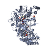
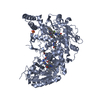
- Structure visualization
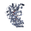
| Structure viewer | Molecule: MolmilJmol/JSmol |
|---|
- Downloads & links
-Download
| PDBx/mmCIF format | 1m8d.cif.gz | 195 KB | Display | PDBx/mmCIF format |
|---|---|---|---|---|
| PDB format | pdb1m8d.ent.gz | 152.5 KB | Display | PDB format |
| PDBx/mmJSON format | 1m8d.json.gz | Tree view | PDBx/mmJSON format | |
| Others |  Other downloads Other downloads |
-Validation report
| Arichive directory | https://data.pdbj.org/pub/pdb/validation_reports/m8/1m8dftp://data.pdbj.org/pub/pdb/validation_reports/m8/1m8d | HTTPS FTP |
|---|
-Related structure data
| Related structure data | 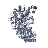 1m8eC 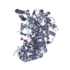 1m8hC  1m8iC  1m9jC 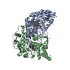 1m9kC 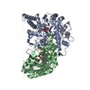 1m9mC 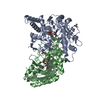 1m9qC 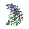 1m9rC 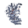 1m9tC 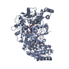 1df1S C: citing same article ( S: Starting model for refinement |
|---|---|
| Similar structure data |


-Links
 PDBj
PDBj


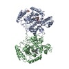
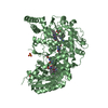
- Assembly
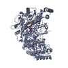
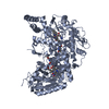
| Deposited unit | 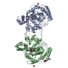
| ||||||||||||||||||||||||||||||
|---|---|---|---|---|---|---|---|---|---|---|---|---|---|---|---|---|---|---|---|---|---|---|---|---|---|---|---|---|---|---|---|
| 1 | 
| ||||||||||||||||||||||||||||||
| 2 | 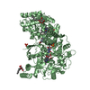
| ||||||||||||||||||||||||||||||
| Unit cell |
| ||||||||||||||||||||||||||||||
| Components on special symmetry positions |
| ||||||||||||||||||||||||||||||
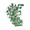
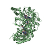
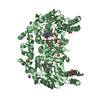
| Details | The biological active assembly is a dimer. There are two monomers in the asymmetric unit, each monomer generates an active dimer. To get one dimer, apply the following symmetry operation to chain A: 1+y-x, y, 1/2-z. / To get a second dimer, apply the following symmetry operation to chain B: 2-x, 1-x+y, 2/3-z+5/3 |
-Components
-Protein / Sugars , 2 types, 3 molecules AB
| #1: Protein | Mass: 50208.027 Da / Num. of mol.: 2 / Fragment: Oxygenase domain Source method: isolated from a genetically manipulated source Source: (gene. exp.)  #6: Sugar | ChemComp-BOG / |  Type: D-saccharide / Mass: 292.369 Da / Num. of mol.: 1 Type: D-saccharide / Mass: 292.369 Da / Num. of mol.: 1Source method: isolated from a genetically manipulated source Formula: C14H28O6 / Comment: detergent*YM |
|---|
-Non-polymers , 6 types, 341 molecules 

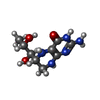
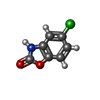


| #2: Chemical |  Mass: 96.063 Da / Num. of mol.: 2 / Source method: obtained synthetically / Formula: SO4 Mass: 96.063 Da / Num. of mol.: 2 / Source method: obtained synthetically / Formula: SO4#3: Chemical |  Mass: 616.487 Da / Num. of mol.: 2 / Source method: obtained synthetically / Formula: C34H32FeN4O4 Mass: 616.487 Da / Num. of mol.: 2 / Source method: obtained synthetically / Formula: C34H32FeN4O4#4: Chemical |  Mass: 241.247 Da / Num. of mol.: 2 / Source method: obtained synthetically / Formula: C9H15N5O3 Mass: 241.247 Da / Num. of mol.: 2 / Source method: obtained synthetically / Formula: C9H15N5O3#5: Chemical |  Mass: 169.565 Da / Num. of mol.: 2 / Source method: obtained synthetically / Formula: C7H4ClNO2 Mass: 169.565 Da / Num. of mol.: 2 / Source method: obtained synthetically / Formula: C7H4ClNO2#7: Chemical | ChemComp-EDO / |  Mass: 62.068 Da / Num. of mol.: 1 / Source method: obtained synthetically / Formula: C2H6O2 Mass: 62.068 Da / Num. of mol.: 1 / Source method: obtained synthetically / Formula: C2H6O2#8: Water | ChemComp-HOH / | Mass: 18.015 Da / Num. of mol.: 332 / Source method: isolated from a natural source / Formula: H2O |
|---|
-Experimental details
-Experiment
| Experiment | Method: X-RAY DIFFRACTION / Number of used crystals: 1 |
|---|
- Sample preparation
| Crystal | Density Matthews: 3.8 Å3/Da / Density % sol: 67.59 % | ||||||||||||||||||||||||||||||||||||||||||||||||||||||||
|---|---|---|---|---|---|---|---|---|---|---|---|---|---|---|---|---|---|---|---|---|---|---|---|---|---|---|---|---|---|---|---|---|---|---|---|---|---|---|---|---|---|---|---|---|---|---|---|---|---|---|---|---|---|---|---|---|---|
| Crystal grow | Temperature: 277 K / Method: vapor diffusion, hanging drop / pH: 6 Details: Lithium Sulfate, B-octyl-glucodise, MES buffer, pH 6.0, VAPOR DIFFUSION, HANGING DROP, temperature 277K | ||||||||||||||||||||||||||||||||||||||||||||||||||||||||
| Crystal grow | *PLUS Temperature: 4 ℃ / pH: 7.6 / Method: vapor diffusion | ||||||||||||||||||||||||||||||||||||||||||||||||||||||||
| Components of the solutions | *PLUS
|
-Data collection
| Diffraction | Mean temperature: 100 K |
|---|---|
| Diffraction source | Source: SYNCHROTRON / Site: SSRL  / Beamline: BL7-1 / Wavelength: 1.08 Å / Beamline: BL7-1 / Wavelength: 1.08 Å |
| Detector | Type: MARRESEARCH / Detector: IMAGE PLATE / Date: Dec 12, 1999 |
| Radiation | Protocol: SINGLE WAVELENGTH / Monochromatic (M) / Laue (L): M / Scattering type: x-ray |
| Radiation wavelength | Wavelength: 1.08 Å / Relative weight: 1 |
| Reflection | Resolution: 2.35→20 Å / Num. obs: 61961 / % possible obs: 95.6 % / Observed criterion σ(F): 0 / Observed criterion σ(I): 0 / Redundancy: 4.96 % / Biso Wilson estimate: 42.1 Å2 / Rmerge(I) obs: 0.058 / Net I/σ(I): 24.8 |
| Reflection shell | Resolution: 2.35→2.43 Å / Redundancy: 2.7 % / Rmerge(I) obs: 0.366 / Mean I/σ(I) obs: 2.5 / Num. unique all: 5149 / Rsym value: 0.366 / % possible all: 80.8 |
| Reflection | *PLUS Num. measured all: 306715 |
| Reflection shell | *PLUS % possible obs: 80.8 % / Num. unique obs: 5149 / Num. measured obs: 13672 |
- Processing
| Software |
| ||||||||||||||||||||||||||||||||||||||||||||||||||||||||||||||||||||||||||||||||
|---|---|---|---|---|---|---|---|---|---|---|---|---|---|---|---|---|---|---|---|---|---|---|---|---|---|---|---|---|---|---|---|---|---|---|---|---|---|---|---|---|---|---|---|---|---|---|---|---|---|---|---|---|---|---|---|---|---|---|---|---|---|---|---|---|---|---|---|---|---|---|---|---|---|---|---|---|---|---|---|---|---|
| Refinement | Method to determine structure: MOLECULAR REPLACEMENT Starting model: PDB entry 1DF1 Resolution: 2.35→19.84 Å / Rfactor Rfree error: 0.005 / Isotropic thermal model: RESTRAINED / Cross valid method: THROUGHOUT / σ(F): 0 / Stereochemistry target values: Engh & Huber
| ||||||||||||||||||||||||||||||||||||||||||||||||||||||||||||||||||||||||||||||||
| Solvent computation | Solvent model: FLAT MODEL / Bsol: 30.321 Å2 / ksol: 0.303507 e/Å3 | ||||||||||||||||||||||||||||||||||||||||||||||||||||||||||||||||||||||||||||||||
| Displacement parameters | Biso mean: 56.3 Å2
| ||||||||||||||||||||||||||||||||||||||||||||||||||||||||||||||||||||||||||||||||
| Refine analyze | Luzzati coordinate error free: 0.39 Å / Luzzati sigma a free: 0.4 Å | ||||||||||||||||||||||||||||||||||||||||||||||||||||||||||||||||||||||||||||||||
| Refinement step | Cycle: LAST / Resolution: 2.35→19.84 Å
| ||||||||||||||||||||||||||||||||||||||||||||||||||||||||||||||||||||||||||||||||
| Refine LS restraints |
| ||||||||||||||||||||||||||||||||||||||||||||||||||||||||||||||||||||||||||||||||
| Refine LS restraints NCS | NCS model details: CONSTR | ||||||||||||||||||||||||||||||||||||||||||||||||||||||||||||||||||||||||||||||||
| LS refinement shell | Resolution: 2.35→2.5 Å / Rfactor Rfree error: 0.018 / Total num. of bins used: 6
| ||||||||||||||||||||||||||||||||||||||||||||||||||||||||||||||||||||||||||||||||
| Xplor file |
| ||||||||||||||||||||||||||||||||||||||||||||||||||||||||||||||||||||||||||||||||
| Refinement | *PLUS Lowest resolution: 20 Å / Rfactor Rfree: 0.274 / Rfactor Rwork: 0.245 | ||||||||||||||||||||||||||||||||||||||||||||||||||||||||||||||||||||||||||||||||
| Solvent computation | *PLUS | ||||||||||||||||||||||||||||||||||||||||||||||||||||||||||||||||||||||||||||||||
| Displacement parameters | *PLUS | ||||||||||||||||||||||||||||||||||||||||||||||||||||||||||||||||||||||||||||||||
| Refine LS restraints | *PLUS
|
