 Movie
Movie Controller

[English] 日本語
 Yorodumi
- PDB-1m5h: Formylmethanofuran:tetrahydromethanopterin formyltransferase from... -
+ Open data

- Basic information
| Entry | Database: PDB / ID: 1m5h | ||||||
|---|---|---|---|---|---|---|---|
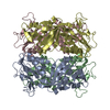
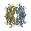
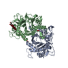
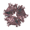
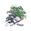
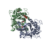
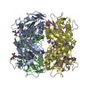
| Title | Formylmethanofuran:tetrahydromethanopterin formyltransferase from Archaeoglobus fulgidus | ||||||
 Components Components | Formylmethanofuran--tetrahydromethanopterin formyltransferase | ||||||
 Keywords Keywords | TRANSFERASE / ALPHA/BETA SANDWICH | ||||||
| Function / homology |  Function and homology information Function and homology informationformylmethanofuran-tetrahydromethanopterin N-formyltransferase / formylmethanofuran-tetrahydromethanopterin N-formyltransferase activity / lactate metabolic process / one-carbon metabolic process / cytoplasm Similarity search - Function | ||||||
| Biological species |   Archaeoglobus fulgidus (archaea) Archaeoglobus fulgidus (archaea) | ||||||
| Method |  X-RAY DIFFRACTION / SYNCHROTRON / MOLECULAR REPLACEMENT / Resolution: 2 Å X-RAY DIFFRACTION / SYNCHROTRON / MOLECULAR REPLACEMENT / Resolution: 2 Å | ||||||
 Authors Authors | Mamat, B. / Roth, A. / Grimm, C. / Ermler, U. / Tziatzios, C. / Schubert, D. / Thauer, R.K. / Shima, S. | ||||||
 Citation Citation | Journal: Protein Sci. / Year: 2002 Title: Crystal structures and enzymatic properties of three formyltransferases from archaea: environmental adaptation and evolutionary relationship. Authors: Mamat, B. / Roth, A. / Grimm, C. / Ermler, U. / Tziatzios, C. / Schubert, D. / Thauer, R.K. / Shima, S. | ||||||
| History |
|
- Structure visualization
| Structure viewer | Molecule: MolmilJmol/JSmol |
|---|
- Downloads & links
-Download
| PDBx/mmCIF format | 1m5h.cif.gz | 489.6 KB | Display | PDBx/mmCIF format |
|---|---|---|---|---|
| PDB format | pdb1m5h.ent.gz | 394.4 KB | Display | PDB format |
| PDBx/mmJSON format | 1m5h.json.gz | Tree view | PDBx/mmJSON format | |
| Others |  Other downloads Other downloads |
-Validation report
| Arichive directory | https://data.pdbj.org/pub/pdb/validation_reports/m5/1m5hftp://data.pdbj.org/pub/pdb/validation_reports/m5/1m5h | HTTPS FTP |
|---|
-Related structure data


-Links
 PDBj
PDBj- Assembly
| Deposited unit | 
| ||||||||||
|---|---|---|---|---|---|---|---|---|---|---|---|
| 1 | 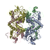
| ||||||||||
| 2 | 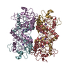
| ||||||||||
| Unit cell |
|
-Components
| #1: Protein | Mass: 31702.188 Da / Num. of mol.: 8 Source method: isolated from a genetically manipulated source Source: (gene. exp.) Archaeoglobus fulgidus (archaea) / Plasmid: pET24b(+) / Species (production host): Escherichia coli / Production host:  References: UniProt: O28076, formylmethanofuran-tetrahydromethanopterin N-formyltransferase #2: Chemical | ChemComp-K /   Mass: 39.098 Da / Num. of mol.: 24 / Source method: obtained synthetically / Formula: K Mass: 39.098 Da / Num. of mol.: 24 / Source method: obtained synthetically / Formula: K#3: Water | ChemComp-HOH / |  Mass: 18.015 Da / Num. of mol.: 1794 / Source method: isolated from a natural source / Formula: H2O Mass: 18.015 Da / Num. of mol.: 1794 / Source method: isolated from a natural source / Formula: H2O |
|---|
-Experimental details
-Experiment
| Experiment | Method: X-RAY DIFFRACTION / Number of used crystals: 1 |
|---|
- Sample preparation
| Crystal | Density Matthews: 2.31 Å3/Da / Density % sol: 46.72 % | ||||||||||||||||||||||||||||||||||||
|---|---|---|---|---|---|---|---|---|---|---|---|---|---|---|---|---|---|---|---|---|---|---|---|---|---|---|---|---|---|---|---|---|---|---|---|---|---|
| Crystal grow | Temperature: 277 K / Method: vapor diffusion, hanging drop / pH: 7 Details: Imidazol/Malate, KSCN, PEG 3350, pH 7.0, VAPOR DIFFUSION, HANGING DROP at 277K | ||||||||||||||||||||||||||||||||||||
| Crystal grow | *PLUS Temperature: 4 ℃ | ||||||||||||||||||||||||||||||||||||
| Components of the solutions | *PLUS
|
-Data collection
| Diffraction | Mean temperature: 100 K |
|---|---|
| Diffraction source | Source: SYNCHROTRON / Site: EMBL/DESY, HAMBURG  / Beamline: BW7B / Wavelength: 0.8452 Å / Beamline: BW7B / Wavelength: 0.8452 Å |
| Detector | Type: MARRESEARCH / Detector: IMAGE PLATE / Date: Jun 20, 2001 |
| Radiation | Protocol: SINGLE WAVELENGTH / Monochromatic (M) / Laue (L): M / Scattering type: x-ray |
| Radiation wavelength | Wavelength: 0.8452 Å / Relative weight: 1 |
| Reflection | Resolution: 2→30 Å / Num. all: 175340 / Num. obs: 150792 / % possible obs: 86 % / Observed criterion σ(I): 1 / Biso Wilson estimate: 5.2 Å2 / Rsym value: 0.08 |
| Reflection shell | Resolution: 2→2.1 Å / Rsym value: 0.223 / % possible all: 67 |
| Reflection | *PLUS Lowest resolution: 30 Å / % possible obs: 86 % / Num. measured all: 839340 / Rmerge(I) obs: 0.08 |
| Reflection shell | *PLUS % possible obs: 67 % / Rmerge(I) obs: 0.223 |
- Processing
| Software |
| ||||||||||||||||||||||||||||||||||||
|---|---|---|---|---|---|---|---|---|---|---|---|---|---|---|---|---|---|---|---|---|---|---|---|---|---|---|---|---|---|---|---|---|---|---|---|---|---|
| Refinement | Method to determine structure: MOLECULAR REPLACEMENT Starting model: Formylmethanofuran:Tetrahydromethanopterin Formyltransferase from Methanosarcina barkeri Resolution: 2→29.25 Å / Rfactor Rfree error: 0.004 / Isotropic thermal model: RESTRAINED / Cross valid method: THROUGHOUT / σ(F): 0 / Stereochemistry target values: Engh & Huber / Details: BULK SOLVENT MODEL USED
| ||||||||||||||||||||||||||||||||||||
| Solvent computation | Solvent model: FLAT MODEL / Bsol: 65.8486 Å2 / ksol: 0.346027 e/Å3 | ||||||||||||||||||||||||||||||||||||
| Displacement parameters | Biso mean: 18 Å2
| ||||||||||||||||||||||||||||||||||||
| Refine analyze | Luzzati coordinate error free: 0.33 Å / Luzzati sigma a free: 0.25 Å | ||||||||||||||||||||||||||||||||||||
| Refinement step | Cycle: LAST / Resolution: 2→29.25 Å
| ||||||||||||||||||||||||||||||||||||
| Refine LS restraints |
| ||||||||||||||||||||||||||||||||||||
| LS refinement shell | Resolution: 2→2.13 Å / Rfactor Rfree error: 0.011 / Total num. of bins used: 6
| ||||||||||||||||||||||||||||||||||||
| Xplor file |
| ||||||||||||||||||||||||||||||||||||
| Refinement | *PLUS % reflection Rfree: 5 % / Rfactor all: 0.229 / Rfactor Rfree: 0.282 / Rfactor Rwork: 0.228 | ||||||||||||||||||||||||||||||||||||
| Solvent computation | *PLUS | ||||||||||||||||||||||||||||||||||||
| Displacement parameters | *PLUS | ||||||||||||||||||||||||||||||||||||
| Refine LS restraints | *PLUS
| ||||||||||||||||||||||||||||||||||||
| LS refinement shell | *PLUS Rfactor Rfree: 0.31 / Rfactor Rwork: 0.248 |
