 ムービー
ムービー コントローラー


+ データを開く

- 基本情報
基本情報
| 登録情報 | データベース: PDB / ID: 1m4x | ||||||
|---|---|---|---|---|---|---|---|
| タイトル | PBCV-1 virus capsid, quasi-atomic model | ||||||
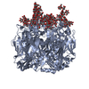
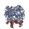
 要素 要素 | PBCV-1 virus capsid | ||||||
 キーワード キーワード | VIRUS / icosahedral virus capsid / beta barrel / Icosahedral virus | ||||||
| 機能・相同性 |  機能・相同性情報 機能・相同性情報 | ||||||
| 生物種 |  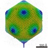 Paramecium bursaria Chlorella virus 1 (ウイルス) Paramecium bursaria Chlorella virus 1 (ウイルス) | ||||||
| 手法 | 電子顕微鏡法 / 単粒子再構成法 / クライオ電子顕微鏡法 / 解像度: 28 Å | ||||||
 データ登録者 データ登録者 | Nandhagopal, N. / Simpson, A.A. / Gurnon, J.R. / Yan, X. / Baker, T.S. / Graves, M.V. / Van Etten, J.L. / Rossmann, M.G. | ||||||
 引用 引用 | ジャーナル: Proc Natl Acad Sci U S A / 年: 2002 タイトル: The structure and evolution of the major capsid protein of a large, lipid-containing DNA virus. 著者: Narayanasamy Nandhagopal / Alan A Simpson / James R Gurnon / Xiadong Yan / Timothy S Baker / Michael V Graves / James L Van Etten / Michael G Rossmann /  要旨: Paramecium bursaria Chlorella virus type 1 (PBCV-1) is a very large, icosahedral virus containing an internal membrane enclosed within a glycoprotein coat consisting of pseudohexagonal arrays of ...Paramecium bursaria Chlorella virus type 1 (PBCV-1) is a very large, icosahedral virus containing an internal membrane enclosed within a glycoprotein coat consisting of pseudohexagonal arrays of trimeric capsomers. Each capsomer is composed of three molecules of the major capsid protein, Vp54, the 2.0-A resolution structure of which is reported here. Four N-linked and two O-linked glycosylation sites were identified. The N-linked sites are associated with nonstandard amino acid motifs as a result of glycosylation by virus-encoded enzymes. Each monomer of the trimeric structure consists of two eight-stranded, antiparallel beta-barrel, "jelly-roll" domains related by a pseudo-sixfold rotation. The fold of the monomer and the pseudo-sixfold symmetry of the capsomer resembles that of the major coat proteins in the double-stranded DNA bacteriophage PRD1 and the double-stranded DNA human adenoviruses, as well as the viral proteins VP2-VP3 of picornaviruses. The structural similarities among these diverse groups of viruses, whose hosts include bacteria, unicellular eukaryotes, plants, and mammals, make it probable that their capsid proteins have evolved from a common ancestor that had already acquired a pseudo-sixfold organization. The trimeric capsid protein structure was used to produce a quasi-atomic model of the 1,900-A diameter PBCV-1 outer shell, based on fitting of the Vp54 crystal structure into a three-dimensional cryoelectron microscopy image reconstruction of the virus. | ||||||
| 履歴 |
|
- 構造の表示
構造の表示
| ムービー |
ムービービューア |
|---|---|
| 構造ビューア | 分子: MolmilJmol/JSmol |
- ダウンロードとリンク
-ダウンロード
| PDBx/mmCIF形式 | 1m4x.cif.gz | 242.1 KB | 表示 | PDBx/mmCIF形式 |
|---|---|---|---|---|
| PDB形式 | pdb1m4x.ent.gz | 275.6 KB | 表示 | PDB形式 |
| PDBx/mmJSON形式 | 1m4x.json.gz | ツリー表示 | PDBx/mmJSON形式 | |
| その他 |  その他のダウンロード その他のダウンロード |
-検証レポート
| アーカイブディレクトリ | https://data.pdbj.org/pub/pdb/validation_reports/m4/1m4xftp://data.pdbj.org/pub/pdb/validation_reports/m4/1m4x | HTTPS FTP |
|---|
-関連構造データ


-リンク
 PDBj
PDBj

- 集合体
集合体
| 登録構造単位 | 
|
|---|---|
| 1 | x 1680
|
| 2 | x 28
|
| 3 | x 140
|
| 4 | x 168
|
| 5 | x 30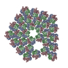
|
| 6 | x 66
|
| 7 | x 28
|
| 対称性 | 点対称性: (ヘルマン・モーガン記号: 532 / シェーンフリース記号: I (正20面体型対称)) |
-要素
| #1: タンパク質 | 分子量: 45734.891 Da / 分子数: 3 / 断片: virus capsid / 由来タイプ: 天然 詳細: Virus infects chlorella algae, which are symbionts with Paramecium bursaria 由来: (天然) Paramecium bursaria Chlorella virus 1 (ウイルス)属: Chlorovirus / 参照: GenBank: 323324, UniProt: P30328*PLUS |
|---|
-実験情報
-実験
| 実験 | 手法: 電子顕微鏡法 |
|---|---|
| EM実験 | 試料の集合状態: PARTICLE / 3次元再構成法: 単粒子再構成法 |
- 試料調製
試料調製
| 構成要素 | 名称: PBCV-1 virus capsid / タイプ: VIRUS / 詳細: T=169d quasi-symmetric icosahedron. |
|---|---|
| ウイルスについての詳細 | ホストのカテゴリ: ALGAE / タイプ: VIRION |
| 緩衝液 | pH: 7 |
| 試料 | 包埋: NO / シャドウイング: NO / 染色: NO / 凍結: YES |
| 試料支持 | 詳細: electron microscopy grid - in vitrious ice |
| 急速凍結 | 詳細: frozen in liquid propane |
| 結晶化 | *PLUS 手法: cryo electron microscopy |
- 電子顕微鏡撮影
| 顕微鏡 | モデル: FEI/PHILIPS CM200FEG/ST |
|---|---|
| 電子銃 | 電子線源:  FIELD EMISSION GUN / 加速電圧: 200 kV / 照射モード: FLOOD BEAM FIELD EMISSION GUN / 加速電圧: 200 kV / 照射モード: FLOOD BEAM |
| 電子レンズ | モード: BRIGHT FIELD |
| 試料ホルダ | 温度: 100 K / 傾斜角・最大: 0 ° / 傾斜角・最小: 0 ° |
| 撮影 | フィルム・検出器のモデル: KODAK SO-163 FILM |
- 解析
解析
| EMソフトウェア |
| ||||||||||||
|---|---|---|---|---|---|---|---|---|---|---|---|---|---|
| CTF補正 | 詳細: inverse of CTF was applied to images | ||||||||||||
| 対称性 | 点対称性: I (正20面体型対称) | ||||||||||||
| 3次元再構成 | 手法: polar fourier transform. / 解像度: 28 Å / 詳細: used Tim Baker's programs PFT, EM3DR, etc. / 対称性のタイプ: POINT | ||||||||||||
| 原子モデル構築 | プロトコル: RIGID BODY FIT / 空間: REAL Target criteria: rigid body refinement in real space against Lagrangian filtered EM density, using the program SITUS.Each molecule in the icosahedral ASU was refined separately. 詳細: METHOD--6d search, separately for each symmetry related molecule in the icosahedral ASU. REFINEMENT PROTOCOL--rigid body | ||||||||||||
| 原子モデル構築 | 3D fitting-ID: 1 / 詳細: 1J5Q or 1M3Y (not exactly as found in pdb entry) / Source name: PDB / タイプ: experimental model
| ||||||||||||
| 精密化ステップ | サイクル: LAST
|

