Major capsid protein, N-terminal / Major capsid protein N-terminus / Major capsid protein, C-terminal / Major capsid protein, C-terminal domain superfamily / Large eukaryotic DNA virus major capsid protein / Group II dsDNA virus coat/capsid protein Similarity search - Domain/homology
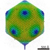
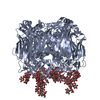
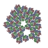
Journal: Proc Natl Acad Sci U S A / Year: 2002 Title: The structure and evolution of the major capsid protein of a large, lipid-containing DNA virus. Authors: Narayanasamy Nandhagopal / Alan A Simpson / James R Gurnon / Xiadong Yan / Timothy S Baker / Michael V Graves / James L Van Etten / Michael G Rossmann / Abstract: Paramecium bursaria Chlorella virus type 1 (PBCV-1) is a very large, icosahedral virus containing an internal membrane enclosed within a glycoprotein coat consisting of pseudohexagonal arrays of ...Paramecium bursaria Chlorella virus type 1 (PBCV-1) is a very large, icosahedral virus containing an internal membrane enclosed within a glycoprotein coat consisting of pseudohexagonal arrays of trimeric capsomers. Each capsomer is composed of three molecules of the major capsid protein, Vp54, the 2.0-A resolution structure of which is reported here. Four N-linked and two O-linked glycosylation sites were identified. The N-linked sites are associated with nonstandard amino acid motifs as a result of glycosylation by virus-encoded enzymes. Each monomer of the trimeric structure consists of two eight-stranded, antiparallel beta-barrel, "jelly-roll" domains related by a pseudo-sixfold rotation. The fold of the monomer and the pseudo-sixfold symmetry of the capsomer resembles that of the major coat proteins in the double-stranded DNA bacteriophage PRD1 and the double-stranded DNA human adenoviruses, as well as the viral proteins VP2-VP3 of picornaviruses. The structural similarities among these diverse groups of viruses, whose hosts include bacteria, unicellular eukaryotes, plants, and mammals, make it probable that their capsid proteins have evolved from a common ancestor that had already acquired a pseudo-sixfold organization. The trimeric capsid protein structure was used to produce a quasi-atomic model of the 1,900-A diameter PBCV-1 outer shell, based on fitting of the Vp54 crystal structure into a three-dimensional cryoelectron microscopy image reconstruction of the virus.
Method: polar fourier transform. / Resolution: 28 Å / Details: used Tim Baker's programs PFT, EM3DR, etc. / Symmetry type: POINT
Atomic model building
Protocol: RIGID BODY FIT / Space: REAL Target criteria: rigid body refinement in real space against Lagrangian filtered EM density, using the program SITUS.Each molecule in the icosahedral ASU was refined separately. Details: METHOD--6d search, separately for each symmetry related molecule in the icosahedral ASU. REFINEMENT PROTOCOL--rigid body
Atomic model building
3D fitting-ID: 1 / Details: 1J5Q or 1M3Y (not exactly as found in pdb entry) / Source name: PDB / Type: experimental model
In the structure databanks used in Yorodumi, some data are registered as the other names, "COVID-19 virus" and "2019-nCoV". Here are the details of the virus and the list of structure data.
Jan 31, 2019. EMDB accession codes are about to change! (news from PDBe EMDB page)
EMDB accession codes are about to change! (news from PDBe EMDB page)
The allocation of 4 digits for EMDB accession codes will soon come to an end. Whilst these codes will remain in use, new EMDB accession codes will include an additional digit and will expand incrementally as the available range of codes is exhausted. The current 4-digit format prefixed with “EMD-” (i.e. EMD-XXXX) will advance to a 5-digit format (i.e. EMD-XXXXX), and so on. It is currently estimated that the 4-digit codes will be depleted around Spring 2019, at which point the 5-digit format will come into force.
The EM Navigator/Yorodumi systems omit the EMD- prefix.
Related info.:Q: What is EMD? / ID/Accession-code notation in Yorodumi/EM Navigator
Yorodumi is a browser for structure data from EMDB, PDB, SASBDB, etc.
This page is also the successor to EM Navigator detail page, and also detail information page/front-end page for Omokage search.
The word "yorodu" (or yorozu) is an old Japanese word meaning "ten thousand". "mi" (miru) is to see.
Related info.:EMDB / PDB / SASBDB / Comparison of 3 databanks / Yorodumi Search / Aug 31, 2016. New EM Navigator & Yorodumi / Yorodumi Papers / Jmol/JSmol / Function and homology information / Changes in new EM Navigator and Yorodumi
 Movie
Movie Controller


 Open data
 Basic information
 Components
 Keywords
 Function and homology information
 Paramecium bursaria Chlorella virus 1
 Authors
 Citation
 Structure visualization
 Downloads & links
 Other downloads
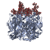


 PDBj
PDBj

 Assembly


 Sample preparation
 Electron microscopy imaging
 FIELD EMISSION GUN / Accelerating voltage: 200 kV / Illumination mode: FLOOD BEAM
 Processing
