 Movie
Movie Controller

[English] 日本語
 Yorodumi
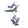
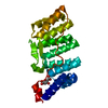
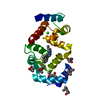
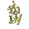
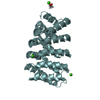
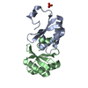
- PDB-1m45: CRYSTAL STRUCTURE OF MLC1P BOUND TO IQ2 OF MYO2P, A CLASS V MYOSIN -
+ Open data

- Basic information
| Entry | Database: PDB / ID: 1m45 | ||||||
|---|---|---|---|---|---|---|---|
| Title | CRYSTAL STRUCTURE OF MLC1P BOUND TO IQ2 OF MYO2P, A CLASS V MYOSIN | ||||||
 Components Components |
| ||||||
 Keywords Keywords | CELL CYCLE PROTEIN / Protein-Peptide complex / IQ motif / myosin light chain | ||||||
| Function / homology |  Function and homology information Function and homology informationprotein localization to cell division site involved in mitotic actomyosin contractile ring assembly / MIH complex / RHO GTPases activate PAKs / regulation of actomyosin contractile ring contraction / peroxisome inheritance / regulation of cell wall organization or biogenesis / myosin II heavy chain binding / RHOT2 GTPase cycle / RHOT1 GTPase cycle / Myo2p-Vac17p-Vac8p transport complex ...protein localization to cell division site involved in mitotic actomyosin contractile ring assembly / MIH complex / RHO GTPases activate PAKs / regulation of actomyosin contractile ring contraction / peroxisome inheritance / regulation of cell wall organization or biogenesis / myosin II heavy chain binding / RHOT2 GTPase cycle / RHOT1 GTPase cycle / Myo2p-Vac17p-Vac8p transport complex / membrane addition at site of cytokinesis / mitochondrion inheritance / site of polarized growth / mitotic actomyosin contractile ring assembly / RHOU GTPase cycle / meiotic nuclear membrane microtubule tethering complex / cellular bud neck contractile ring / vesicle targeting / myosin V complex / vacuole inheritance / vesicle transport along actin filament / incipient cellular bud site / septum digestion after cytokinesis / cellular bud tip / Golgi inheritance / myosin V binding / cellular bud neck / mating projection tip / fungal-type vacuole membrane / vesicle docking involved in exocytosis / myosin II complex / microfilament motor activity / actin filament bundle / filamentous actin / intracellular distribution of mitochondria / establishment of mitotic spindle orientation / transport vesicle / vesicle-mediated transport / actin filament organization / regulation of cytokinesis / small GTPase binding / actin filament binding / protein transport / actin cytoskeleton / vesicle / calmodulin binding / calcium ion binding / ATP binding / identical protein binding / membrane / plasma membrane / cytoplasm Similarity search - Function | ||||||
| Biological species |  | ||||||
| Method |  X-RAY DIFFRACTION / SYNCHROTRON / MOLECULAR REPLACEMENT, Structure of MLC1P bound to IQ 2, 3 (SOLVED BY MAD METHOD) / Resolution: 1.65 Å X-RAY DIFFRACTION / SYNCHROTRON / MOLECULAR REPLACEMENT, Structure of MLC1P bound to IQ 2, 3 (SOLVED BY MAD METHOD) / Resolution: 1.65 Å | ||||||
 Authors Authors | Terrak, M. / Dominguez, R. | ||||||
 Citation Citation | Journal: Embo J. / Year: 2003 Title: Two distinct myosin light chain structures are induced by specific variations within the bound IQ motifs-functional implications Authors: Terrak, M. / Wu, G. / Stafford, W.F. / Lu, R.C. / Dominguez, R. #1: Journal: Acta Crystallogr.,Sect.D / Year: 2002Title: Crystallisation, X-ray characterization and selenomethionine phasing of Mlc1p bound to IQ motifs from myosin V Authors: Terrak, M. / Otterbein, L.R. / Wu, G. / Palecanda, L.A. / Lu, R.C. / Dominguez, R. | ||||||
| History |
|
- Structure visualization
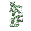
| Structure viewer | Molecule: MolmilJmol/JSmol |
|---|
- Downloads & links
-Download
| PDBx/mmCIF format | 1m45.cif.gz | 51.9 KB | Display | PDBx/mmCIF format |
|---|---|---|---|---|
| PDB format | pdb1m45.ent.gz | 37.5 KB | Display | PDB format |
| PDBx/mmJSON format | 1m45.json.gz | Tree view | PDBx/mmJSON format | |
| Others |  Other downloads Other downloads |
-Validation report
| Summary document | 1m45_validation.pdf.gz | 365.7 KB | Display | wwPDB validaton report |
|---|---|---|---|---|
| Full document | 1m45_full_validation.pdf.gz | 368.3 KB | Display | |
| Data in XML | 1m45_validation.xml.gz | 5 KB | Display | |
| Data in CIF | 1m45_validation.cif.gz | 8.4 KB | Display | |
| Arichive directory | https://data.pdbj.org/pub/pdb/validation_reports/m4/1m45ftp://data.pdbj.org/pub/pdb/validation_reports/m4/1m45 | HTTPS FTP |
-Related structure data

-Links
 PDBj
PDBj


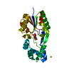
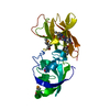
- Assembly
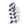
| Deposited unit | 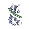
| ||||||||
|---|---|---|---|---|---|---|---|---|---|
| 1 |
| ||||||||
| Unit cell |
|
-Components
| #1: Protein | Mass: 16332.213 Da / Num. of mol.: 1 Source method: isolated from a genetically manipulated source Source: (gene. exp.) Gene: MLC1 / Plasmid: pAED4 / Species (production host): Escherichia coli / Production host:  |
|---|---|
| #2: Protein/peptide | Mass: 2993.420 Da / Num. of mol.: 1 / Source method: obtained synthetically Details: The peptide was chemically synthesized. The sequence of the peptide is naturally found in Saccharomyces cerevisiae (Baker's yeast). References: UniProt: P19524 |
| #3: Water | ChemComp-HOH /  Mass: 18.015 Da / Num. of mol.: 252 / Source method: isolated from a natural source / Formula: H2O Mass: 18.015 Da / Num. of mol.: 252 / Source method: isolated from a natural source / Formula: H2O |
-Experimental details
-Experiment
| Experiment | Method: X-RAY DIFFRACTION / Number of used crystals: 1 |
|---|
- Sample preparation
| Crystal | Density Matthews: 1.81 Å3/Da / Density % sol: 32.13 % |
|---|---|
| Crystal grow | Temperature: 293 K / Method: vapor diffusion, hanging drop Details: PEG 3350, sodium chloride, VAPOR DIFFUSION, HANGING DROP, temperature 293K |
-Data collection
| Diffraction | Mean temperature: 100 K |
|---|---|
| Diffraction source | Source: SYNCHROTRON / Site: APS  / Beamline: 14-BM-D / Wavelength: 1 Å / Beamline: 14-BM-D / Wavelength: 1 Å |
| Detector | Type: ADSC QUANTUM 4 / Detector: CCD / Date: Jun 1, 2000 |
| Radiation | Monochromator: Si(111) double-crystal monochromator / Protocol: SINGLE WAVELENGTH / Monochromatic (M) / Laue (L): M / Scattering type: x-ray |
| Radiation wavelength | Wavelength: 1 Å / Relative weight: 1 |
| Reflection | Resolution: 1.64→25.4 Å / Num. all: 17258 / Num. obs: 17258 / % possible obs: 97.1 % / Observed criterion σ(F): 0 / Observed criterion σ(I): 0 / Redundancy: 10.3 % / Rmerge(I) obs: 0.036 / Net I/σ(I): 25.6 |
| Reflection shell | Resolution: 1.64→1.71 Å / Rmerge(I) obs: 0.2 / Mean I/σ(I) obs: 6.1 / % possible all: 86 |
- Processing
| Software |
| |||||||||||||||||||||||||||||||||||||||||||||||||||||||||||||||||||||||||||
|---|---|---|---|---|---|---|---|---|---|---|---|---|---|---|---|---|---|---|---|---|---|---|---|---|---|---|---|---|---|---|---|---|---|---|---|---|---|---|---|---|---|---|---|---|---|---|---|---|---|---|---|---|---|---|---|---|---|---|---|---|---|---|---|---|---|---|---|---|---|---|---|---|---|---|---|---|
| Refinement | Method to determine structure: MOLECULAR REPLACEMENT, Structure of MLC1P bound to IQ 2, 3 (SOLVED BY MAD METHOD) Resolution: 1.65→25 Å / Cor.coef. Fo:Fc: 0.952 / Cor.coef. Fo:Fc free: 0.936 / SU B: 2.229 / SU ML: 0.076 / Isotropic thermal model: overall anisotropic / Cross valid method: THROUGHOUT / σ(F): 0 / ESU R: 0.131 / ESU R Free: 0.121 / Stereochemistry target values: MAXIMUM LIKELIHOOD Details: CNS 1.0 was also used at the beginning of the refinement of this structure
| |||||||||||||||||||||||||||||||||||||||||||||||||||||||||||||||||||||||||||
| Solvent computation | Ion probe radii: 0.8 Å / Shrinkage radii: 0.8 Å / VDW probe radii: 1.4 Å / Solvent model: BABINET MODEL WITH MASK | |||||||||||||||||||||||||||||||||||||||||||||||||||||||||||||||||||||||||||
| Displacement parameters | Biso mean: 24.235 Å2
| |||||||||||||||||||||||||||||||||||||||||||||||||||||||||||||||||||||||||||
| Refinement step | Cycle: LAST / Resolution: 1.65→25 Å
| |||||||||||||||||||||||||||||||||||||||||||||||||||||||||||||||||||||||||||
| Refine LS restraints |
| |||||||||||||||||||||||||||||||||||||||||||||||||||||||||||||||||||||||||||
| LS refinement shell | Resolution: 1.65→1.693 Å / Total num. of bins used: 20
|
