 Movie
Movie Controller

[English] 日本語
 Yorodumi
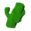
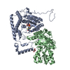
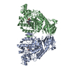
- PDB-1lwj: CRYSTAL STRUCTURE OF T. MARITIMA 4-ALPHA-GLUCANOTRANSFERASE/ACARB... -
+ Open data

- Basic information
| Entry | Database: PDB / ID: 1lwj | ||||||
|---|---|---|---|---|---|---|---|
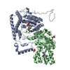
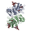
| Title | CRYSTAL STRUCTURE OF T. MARITIMA 4-ALPHA-GLUCANOTRANSFERASE/ACARBOSE COMPLEX | ||||||
 Components Components | 4-ALPHA-GLUCANOTRANSFERASE | ||||||
 Keywords Keywords | TRANSFERASE / 4-alpha-glucanotransferase / alpha-amylase family / Thermotoga maritima / acarbose / (beta/alpha)8 barrel | ||||||
| Function / homology |  Function and homology information Function and homology information4-alpha-glucanotransferase / 4-alpha-glucanotransferase activity / alpha-amylase activity / oligosaccharide catabolic process / metal ion binding / cytoplasm Similarity search - Function | ||||||
| Biological species |   Thermotoga maritima (bacteria) Thermotoga maritima (bacteria) | ||||||
| Method |  X-RAY DIFFRACTION / SYNCHROTRON / MOLECULAR REPLACEMENT / Resolution: 2.5 Å X-RAY DIFFRACTION / SYNCHROTRON / MOLECULAR REPLACEMENT / Resolution: 2.5 Å | ||||||
 Authors Authors | Roujeinikova, A. / Raasch, C. / Sedelnikova, S. / Liebl, W. / Rice, D.W. | ||||||
 Citation Citation | Journal: J.Mol.Biol. / Year: 2002 Title: CRYSTAL STRUCTURE OF THERMOTOGA MARITIMA 4-ALPHA-GLUCANOTRANSFERASE AND ITS ACARBOSE COMPLEX: IMPLICATIONS FOR SUBSTRATE SPECIFICITY AND CATALYSIS Authors: Roujeinikova, A. / Raasch, C. / Sedelnikova, S. / Liebl, W. / Rice, D.W. #1: Journal: Acta Crystallogr.,Sect.D / Year: 2001Title: CRYSTALLIZATION AND PRELIMINARY X-RAY CRYSTALLOGRAPHIC STUDIES ON 4-ALPHA-GLUCANOTRANSFERASE FROM THERMOTOGA MARITIMA Authors: Roujeinikova, A. / Raasch, C. / Sedelnikova, S. / Liebl, W. / Rice, D.W. | ||||||
| History |
|
- Structure visualization
| Structure viewer | Molecule: MolmilJmol/JSmol |
|---|
- Downloads & links
-Download
| PDBx/mmCIF format | 1lwj.cif.gz | 179.3 KB | Display | PDBx/mmCIF format |
|---|---|---|---|---|
| PDB format | pdb1lwj.ent.gz | 143.4 KB | Display | PDB format |
| PDBx/mmJSON format | 1lwj.json.gz | Tree view | PDBx/mmJSON format | |
| Others |  Other downloads Other downloads |
-Validation report
| Arichive directory | https://data.pdbj.org/pub/pdb/validation_reports/lw/1lwjftp://data.pdbj.org/pub/pdb/validation_reports/lw/1lwj | HTTPS FTP |
|---|
-Related structure data
| Related structure data |  1lwhSC S: Starting model for refinement C: citing same article ( |
|---|---|
| Similar structure data |


-Links
 PDBj
PDBj

- Assembly
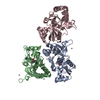
| Deposited unit | 
| ||||||||||||
|---|---|---|---|---|---|---|---|---|---|---|---|---|---|
| 1 |
| ||||||||||||
| Unit cell |
| ||||||||||||
| Components on special symmetry positions |
| ||||||||||||
| Details | The dimer in the asymmetric unit is the biological unit |
-Components
| #1: Protein | Mass: 51925.898 Da / Num. of mol.: 2 Source method: isolated from a genetically manipulated source Source: (gene. exp.) Thermotoga maritima (bacteria) / Gene: mgt / Plasmid: PTAM1 / Production host: #2: Sugar | 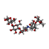  Type: saccharide / Mass: 791.746 Da / Num. of mol.: 2 Type: saccharide / Mass: 791.746 Da / Num. of mol.: 2Source method: isolated from a genetically manipulated source Formula: C31H53NO22 #3: Chemical |   Mass: 40.078 Da / Num. of mol.: 2 / Source method: obtained synthetically / Formula: Ca Mass: 40.078 Da / Num. of mol.: 2 / Source method: obtained synthetically / Formula: Ca#4: Water | ChemComp-HOH / |  Mass: 18.015 Da / Num. of mol.: 41 / Source method: isolated from a natural source / Formula: H2O Mass: 18.015 Da / Num. of mol.: 41 / Source method: isolated from a natural source / Formula: H2O |
|---|
-Experimental details
-Experiment
| Experiment | Method: X-RAY DIFFRACTION / Number of used crystals: 1 |
|---|
- Sample preparation
| Crystal | Density Matthews: 4.07 Å3/Da / Density % sol: 69.78 % |
|---|---|
| Crystal grow | Temperature: 290 K / Method: vapor diffusion, hanging drop / pH: 6.5 Details: 26-29% PEG 5000, 200-300 mM Ammonium Sulphate, 3 mM CaCl2, 80 mM MES, 40 mM acarbose, pH 6.5, VAPOR DIFFUSION, HANGING DROP, temperature 290K |
-Data collection
| Diffraction | Mean temperature: 100 K |
|---|---|
| Diffraction source | Source: SYNCHROTRON / Site: SRS  / Beamline: PX14.2 / Wavelength: 0.978 Å / Beamline: PX14.2 / Wavelength: 0.978 Å |
| Detector | Type: ADSC QUANTUM 4 / Detector: CCD / Date: Feb 11, 2001 |
| Radiation | Monochromator: Triangular single crystal Si monochromator / Protocol: SINGLE WAVELENGTH / Monochromatic (M) / Laue (L): M / Scattering type: x-ray |
| Radiation wavelength | Wavelength: 0.978 Å / Relative weight: 1 |
| Reflection | Resolution: 2.5→15 Å / Num. obs: 39305 / % possible obs: 67 % / Observed criterion σ(I): -3 / Redundancy: 4.8 % / Biso Wilson estimate: 30 Å2 / Rmerge(I) obs: 0.11 / Net I/σ(I): 12.1 |
| Reflection shell | Resolution: 2.5→2.56 Å / Redundancy: 4.2 % / Rmerge(I) obs: 0.22 / Mean I/σ(I) obs: 5.4 / Num. unique all: 979 / % possible all: 25 |
- Processing
| Software |
| ||||||||||||||||||||
|---|---|---|---|---|---|---|---|---|---|---|---|---|---|---|---|---|---|---|---|---|---|
| Refinement | Method to determine structure: MOLECULAR REPLACEMENT Starting model: PDB ENTRY 1LWH Resolution: 2.5→10 Å / Cross valid method: THROUGHOUT / σ(F): 0 / σ(I): 0 / Stereochemistry target values: MLF
| ||||||||||||||||||||
| Refinement step | Cycle: LAST / Resolution: 2.5→10 Å
| ||||||||||||||||||||
| Refine LS restraints |
|
