 Movie
Movie Controller


+ Open data

- Basic information
| Entry | Database: PDB / ID: 1lv7 | ||||||
|---|---|---|---|---|---|---|---|
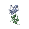
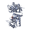
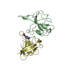
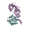
| Title | Crystal Structure of the AAA domain of FtsH | ||||||
 Components Components | FtsH | ||||||
 Keywords Keywords | HYDROLASE / ALPHA/BETA DOMAIN / FOUR HELIX BUNDLE | ||||||
| Function / homology |  Function and homology information Function and homology informationmembrane protein complex / Hydrolases; Acting on peptide bonds (peptidases); Metalloendopeptidases / ATP-dependent peptidase activity / protein catabolic process / metalloendopeptidase activity / ATP hydrolysis activity / proteolysis / zinc ion binding / ATP binding / plasma membrane Similarity search - Function | ||||||
| Biological species |  | ||||||
| Method |  X-RAY DIFFRACTION / SYNCHROTRON / MIRAS / Resolution: 1.5 Å X-RAY DIFFRACTION / SYNCHROTRON / MIRAS / Resolution: 1.5 Å | ||||||
 Authors Authors | Krzywda, S. / Brzozowski, A.M. / Verma, C. / Karata, K. / Ogura, T. / Wilkinson, A.J. | ||||||
 Citation Citation | Journal: Structure / Year: 2002 Title: The crystal structure of the AAA domain of the ATP-dependent protease FtsH of Escherichia coli at 1.5 A resolution. Authors: Krzywda, S. / Brzozowski, A.M. / Verma, C. / Karata, K. / Ogura, T. / Wilkinson, A.J. #1: Journal: Acta Crystallogr.,Sect.D / Year: 2002Title: Crystallization of the AAA domain of the ATP-dependent protease FtsH of Escherichia coli Authors: Krzywda, S. / Brzozowski, A.M. / Karata, K. / Ogura, T. / Wilkinson, A.J. | ||||||
| History |
|
- Structure visualization
| Structure viewer | Molecule: MolmilJmol/JSmol |
|---|
- Downloads & links
-Download
| PDBx/mmCIF format | 1lv7.cif.gz | 124.8 KB | Display | PDBx/mmCIF format |
|---|---|---|---|---|
| PDB format | pdb1lv7.ent.gz | 97.5 KB | Display | PDB format |
| PDBx/mmJSON format | 1lv7.json.gz | Tree view | PDBx/mmJSON format | |
| Others |  Other downloads Other downloads |
-Validation report
| Arichive directory | https://data.pdbj.org/pub/pdb/validation_reports/lv/1lv7ftp://data.pdbj.org/pub/pdb/validation_reports/lv/1lv7 | HTTPS FTP |
|---|
-Related structure data
| Similar structure data |
|---|


-Links
 PDBj
PDBj


- Assembly
| Deposited unit | 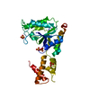
| ||||||||
|---|---|---|---|---|---|---|---|---|---|
| 1 |
| ||||||||
| 2 | 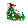
| ||||||||
| Unit cell |
| ||||||||
| Components on special symmetry positions |
|
-Components
| #1: Protein | Mass: 27769.084 Da / Num. of mol.: 1 / Fragment: AAA domain Source method: isolated from a genetically manipulated source Source: (gene. exp.) References: UniProt: P0AAI3, Hydrolases; Acting on peptide bonds (peptidases); Metalloendopeptidases | ||
|---|---|---|---|
| #2: Chemical | ChemComp-SO4 /   Mass: 96.063 Da / Num. of mol.: 5 / Source method: obtained synthetically / Formula: SO4 Mass: 96.063 Da / Num. of mol.: 5 / Source method: obtained synthetically / Formula: SO4#3: Water | ChemComp-HOH / |  Mass: 18.015 Da / Num. of mol.: 366 / Source method: isolated from a natural source / Formula: H2O Mass: 18.015 Da / Num. of mol.: 366 / Source method: isolated from a natural source / Formula: H2O |
-Experimental details
-Experiment
| Experiment | Method: X-RAY DIFFRACTION / Number of used crystals: 1 |
|---|
- Sample preparation
| Crystal | Density Matthews: 2.4 Å3/Da / Density % sol: 48.82 % | |||||||||||||||||||||||||||||||||||||||||||||||||
|---|---|---|---|---|---|---|---|---|---|---|---|---|---|---|---|---|---|---|---|---|---|---|---|---|---|---|---|---|---|---|---|---|---|---|---|---|---|---|---|---|---|---|---|---|---|---|---|---|---|---|
| Crystal grow | Temperature: 292 K / Method: vapor diffusion, hanging drop / pH: 8.5 Details: ammonium sulfate, Tris-HCl, DTT, pH 8.5, VAPOR DIFFUSION, HANGING DROP, temperature 292K | |||||||||||||||||||||||||||||||||||||||||||||||||
| Crystal grow | *PLUS | |||||||||||||||||||||||||||||||||||||||||||||||||
| Components of the solutions | *PLUS
|
-Data collection
| Diffraction | Mean temperature: 120 K |
|---|---|
| Diffraction source | Source: SYNCHROTRON / Site: ESRF  / Beamline: ID14-2 / Wavelength: 0.933 Å / Beamline: ID14-2 / Wavelength: 0.933 Å |
| Detector | Type: ADSC QUANTUM 4 / Detector: CCD / Date: Jan 26, 2002 |
| Radiation | Protocol: SINGLE WAVELENGTH / Monochromatic (M) / Laue (L): M / Scattering type: x-ray |
| Radiation wavelength | Wavelength: 0.933 Å / Relative weight: 1 |
| Reflection | *PLUS Highest resolution: 1.5 Å / Lowest resolution: 50 Å / Num. obs: 44655 / % possible obs: 98.4 % / Num. measured all: 913258 / Rmerge(I) obs: 0.064 |
| Reflection shell | *PLUS Highest resolution: 1.5 Å / Lowest resolution: 1.55 Å / % possible obs: 91.1 % / Rmerge(I) obs: 0.261 / Mean I/σ(I) obs: 2.6 |
- Processing
| Software |
| ||||||||||||||||||||||||||||||||||||||||||||||||||||||||||||||||||||||||||||||||||||||||||||||||||||||||||||||||||||||||||||||||||
|---|---|---|---|---|---|---|---|---|---|---|---|---|---|---|---|---|---|---|---|---|---|---|---|---|---|---|---|---|---|---|---|---|---|---|---|---|---|---|---|---|---|---|---|---|---|---|---|---|---|---|---|---|---|---|---|---|---|---|---|---|---|---|---|---|---|---|---|---|---|---|---|---|---|---|---|---|---|---|---|---|---|---|---|---|---|---|---|---|---|---|---|---|---|---|---|---|---|---|---|---|---|---|---|---|---|---|---|---|---|---|---|---|---|---|---|---|---|---|---|---|---|---|---|---|---|---|---|---|---|---|---|
| Refinement | Method to determine structure: MIRAS / Resolution: 1.5→50 Å / Cor.coef. Fo:Fc: 0.972 / Cor.coef. Fo:Fc free: 0.971 / SU B: 1.129 / SU ML: 0.042 / Cross valid method: THROUGHOUT / ESU R: 0.079 / ESU R Free: 0.065 / Stereochemistry target values: MAXIMUM LIKELIHOOD / Details: HYDROGENS HAVE BEEN ADDED IN THE RIDING POSITIONS
| ||||||||||||||||||||||||||||||||||||||||||||||||||||||||||||||||||||||||||||||||||||||||||||||||||||||||||||||||||||||||||||||||||
| Solvent computation | Ion probe radii: 0.8 Å / Shrinkage radii: 0.8 Å / VDW probe radii: 1.4 Å / Solvent model: BABINET MODEL WITH MASK | ||||||||||||||||||||||||||||||||||||||||||||||||||||||||||||||||||||||||||||||||||||||||||||||||||||||||||||||||||||||||||||||||||
| Displacement parameters | Biso mean: 16.559 Å2
| ||||||||||||||||||||||||||||||||||||||||||||||||||||||||||||||||||||||||||||||||||||||||||||||||||||||||||||||||||||||||||||||||||
| Refinement step | Cycle: LAST / Resolution: 1.5→50 Å
| ||||||||||||||||||||||||||||||||||||||||||||||||||||||||||||||||||||||||||||||||||||||||||||||||||||||||||||||||||||||||||||||||||
| Refine LS restraints |
| ||||||||||||||||||||||||||||||||||||||||||||||||||||||||||||||||||||||||||||||||||||||||||||||||||||||||||||||||||||||||||||||||||
| LS refinement shell | Resolution: 1.5→1.54 Å / Total num. of bins used: 20
| ||||||||||||||||||||||||||||||||||||||||||||||||||||||||||||||||||||||||||||||||||||||||||||||||||||||||||||||||||||||||||||||||||
| Refinement TLS params. | Method: refined / Origin x: 19.9861 Å / Origin y: 21.6216 Å / Origin z: 55.3517 Å
| ||||||||||||||||||||||||||||||||||||||||||||||||||||||||||||||||||||||||||||||||||||||||||||||||||||||||||||||||||||||||||||||||||
| Refinement | *PLUS Highest resolution: 1.5 Å / Lowest resolution: 50 Å / Num. reflection obs: 421544 / % reflection Rfree: 5 % / Rfactor obs: 0.15488 / Rfactor Rfree: 0.178 / Rfactor Rwork: 0.154 | ||||||||||||||||||||||||||||||||||||||||||||||||||||||||||||||||||||||||||||||||||||||||||||||||||||||||||||||||||||||||||||||||||
| Solvent computation | *PLUS | ||||||||||||||||||||||||||||||||||||||||||||||||||||||||||||||||||||||||||||||||||||||||||||||||||||||||||||||||||||||||||||||||||
| Displacement parameters | *PLUS | ||||||||||||||||||||||||||||||||||||||||||||||||||||||||||||||||||||||||||||||||||||||||||||||||||||||||||||||||||||||||||||||||||
| Refine LS restraints | *PLUS
| ||||||||||||||||||||||||||||||||||||||||||||||||||||||||||||||||||||||||||||||||||||||||||||||||||||||||||||||||||||||||||||||||||
| LS refinement shell | *PLUS Rfactor Rfree: 0.239 / Rfactor Rwork: 0.22 |
