 Movie
Movie Controller


+ Open data

- Basic information
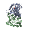
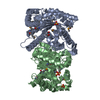
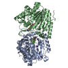
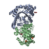
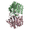
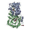
| Entry | Database: PDB / ID: 1luc | ||||||
|---|---|---|---|---|---|---|---|
| Title | BACTERIAL LUCIFERASE | ||||||
 Components Components | (BACTERIAL LUCIFERASE) x 2 | ||||||
 Keywords Keywords | FLAVOPROTEIN / MONOOXYGENASE | ||||||
| Function / homology |  Function and homology information Function and homology informationbacterial luciferase / alkanal monooxygenase (FMN-linked) activity / bioluminescence / cytosol Similarity search - Function | ||||||
| Biological species |  Vibrio harveyi (bacteria) Vibrio harveyi (bacteria) | ||||||
| Method |  X-RAY DIFFRACTION / SYNCHROTRON / Resolution: 1.5 Å X-RAY DIFFRACTION / SYNCHROTRON / Resolution: 1.5 Å | ||||||
 Authors Authors | Fisher, A.J. / Rayment, I. | ||||||
 Citation Citation | Journal: J.Biol.Chem. / Year: 1996 Title: The 1.5-A resolution crystal structure of bacterial luciferase in low salt conditions. Authors: Fisher, A.J. / Thompson, T.B. / Thoden, J.B. / Baldwin, T.O. / Rayment, I. #1: Journal: Biochemistry / Year: 1995Title: Three-Dimensional Structure of Bacterial Luciferase from Vibrio Harveyi at 2.4 A Resolution Authors: Fisher, A.J. / Raushel, F.M. / Baldwin, T.O. / Rayment, I. | ||||||
| History |
|
- Structure visualization
| Structure viewer | Molecule: MolmilJmol/JSmol |
|---|
- Downloads & links
-Download
| PDBx/mmCIF format | 1luc.cif.gz | 155.5 KB | Display | PDBx/mmCIF format |
|---|---|---|---|---|
| PDB format | pdb1luc.ent.gz | 119.1 KB | Display | PDB format |
| PDBx/mmJSON format | 1luc.json.gz | Tree view | PDBx/mmJSON format | |
| Others |  Other downloads Other downloads |
-Validation report
| Summary document | 1luc_validation.pdf.gz | 391.8 KB | Display | wwPDB validaton report |
|---|---|---|---|---|
| Full document | 1luc_full_validation.pdf.gz | 406.7 KB | Display | |
| Data in XML | 1luc_validation.xml.gz | 15.3 KB | Display | |
| Data in CIF | 1luc_validation.cif.gz | 26.6 KB | Display | |
| Arichive directory | https://data.pdbj.org/pub/pdb/validation_reports/lu/1lucftp://data.pdbj.org/pub/pdb/validation_reports/lu/1luc | HTTPS FTP |
-Related structure data
| Similar structure data |
|---|


-Links
 PDBj
PDBj
- Assembly
| Deposited unit | 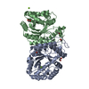
| ||||||||
|---|---|---|---|---|---|---|---|---|---|
| 1 |
| ||||||||
| Unit cell |
|
-Components
| #1: Protein | Mass: 40197.062 Da / Num. of mol.: 1 Source method: isolated from a genetically manipulated source Source: (gene. exp.) Vibrio harveyi (bacteria) / Production host: | ||||
|---|---|---|---|---|---|
| #2: Protein | Mass: 36384.684 Da / Num. of mol.: 1 Source method: isolated from a genetically manipulated source Source: (gene. exp.) Vibrio harveyi (bacteria) / Production host: | ||||
| #3: Chemical |   Mass: 24.305 Da / Num. of mol.: 3 / Source method: obtained synthetically / Formula: Mg Mass: 24.305 Da / Num. of mol.: 3 / Source method: obtained synthetically / Formula: Mg#4: Chemical | ChemComp-EDO /   Mass: 62.068 Da / Num. of mol.: 5 / Source method: obtained synthetically / Formula: C2H6O2 Mass: 62.068 Da / Num. of mol.: 5 / Source method: obtained synthetically / Formula: C2H6O2#5: Water | ChemComp-HOH / |  Mass: 18.015 Da / Num. of mol.: 638 / Source method: isolated from a natural source / Formula: H2O Mass: 18.015 Da / Num. of mol.: 638 / Source method: isolated from a natural source / Formula: H2O |
-Experimental details
-Experiment
| Experiment | Method: X-RAY DIFFRACTION |
|---|
- Sample preparation
| Crystal | Density Matthews: 2.21 Å3/Da / Density % sol: 44 % | ||||||||||||||||||||||||||||||
|---|---|---|---|---|---|---|---|---|---|---|---|---|---|---|---|---|---|---|---|---|---|---|---|---|---|---|---|---|---|---|---|
| Crystal grow | *PLUS Temperature: 4 ℃ / pH: 6.5 / Method: batch method | ||||||||||||||||||||||||||||||
| Components of the solutions | *PLUS
|
-Data collection
| Diffraction source | Source: SYNCHROTRON / Site: SSRL  / Beamline: BL7-1 / Wavelength: 1.08 / Beamline: BL7-1 / Wavelength: 1.08 |
|---|---|
| Detector | Type: MARRESEARCH / Detector: IMAGE PLATE / Date: Jul 1, 1995 |
| Radiation | Monochromatic (M) / Laue (L): M / Scattering type: x-ray |
| Radiation wavelength | Wavelength: 1.08 Å / Relative weight: 1 |
| Reflection | Resolution: 1.5→30 Å / Num. obs: 105012 / Observed criterion σ(I): 0 / Redundancy: 2.8 % / Rmerge(I) obs: 0.041 |
| Reflection | *PLUS Num. obs: 105158 / % possible obs: 99 % / Num. measured all: 297839 |
- Processing
| Software |
| ||||||||||||||||||||||||||||||
|---|---|---|---|---|---|---|---|---|---|---|---|---|---|---|---|---|---|---|---|---|---|---|---|---|---|---|---|---|---|---|---|
| Refinement | Resolution: 1.5→30 Å / σ(F): 0 /
| ||||||||||||||||||||||||||||||
| Displacement parameters | Biso mean: 22.2 Å2 | ||||||||||||||||||||||||||||||
| Refinement step | Cycle: LAST / Resolution: 1.5→30 Å
| ||||||||||||||||||||||||||||||
| Refine LS restraints |
| ||||||||||||||||||||||||||||||
| Software | *PLUS Name: TNT / Classification: refinement | ||||||||||||||||||||||||||||||
| Refinement | *PLUS Num. reflection obs: 105018 / Rfactor obs: 0.182 | ||||||||||||||||||||||||||||||
| Solvent computation | *PLUS | ||||||||||||||||||||||||||||||
| Displacement parameters | *PLUS | ||||||||||||||||||||||||||||||
| Refine LS restraints | *PLUS
|
