 Movie
Movie Controller

[English] 日本語
 Yorodumi
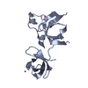
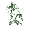
- PDB-1lu0: Atomic Resolution Structure of Squash Trypsin Inhibitor: Unexpect... -
+ Open data

- Basic information
| Entry | Database: PDB / ID: 1lu0 | ||||||
|---|---|---|---|---|---|---|---|
| Title | Atomic Resolution Structure of Squash Trypsin Inhibitor: Unexpected Metal Coordination | ||||||
 Components Components | Trypsin inhibitor I | ||||||
 Keywords Keywords | HYDROLASE INHIBITOR / serine protease inhibitor / metal coordination | ||||||
| Function / homology | Proteinase inhibitor I7, squash / Squash family serine protease inhibitor / Squash family of serine protease inhibitors signature. / Plant trypsin inhibitors / Proteinase/amylase inhibitor domain superfamily / serine-type endopeptidase inhibitor activity / extracellular region / Trypsin inhibitor 1 Function and homology information Function and homology information | ||||||
| Biological species |  Cucurbita maxima (winter squash) Cucurbita maxima (winter squash) | ||||||
| Method |  X-RAY DIFFRACTION / SYNCHROTRON / Dual-space recycling / Resolution: 1.03 Å X-RAY DIFFRACTION / SYNCHROTRON / Dual-space recycling / Resolution: 1.03 Å | ||||||
 Authors Authors | Thaimattam, R. / Tykarska, E. / Bierzynski, A. / Sheldrick, G.M. / Jaskolski, M. | ||||||
 Citation Citation | Journal: Acta Crystallogr.,Sect.D / Year: 2002 Title: Atomic resolution structure of squash trypsin inhibitor: unexpected metal coordination. Authors: Thaimattam, R. / Tykarska, E. / Bierzynski, A. / Sheldrick, G.M. / Jaskolski, M. #1: Journal: FEBS Lett. / Year: 1989Title: The refined 2.0 A X-ray crystal structure of the complex formed between bovine beta-trypsin and CMTI-I, a trypsin inhibitor from squash seeds (Cucurbita maxima). Topological similarity of the ...Title: The refined 2.0 A X-ray crystal structure of the complex formed between bovine beta-trypsin and CMTI-I, a trypsin inhibitor from squash seeds (Cucurbita maxima). Topological similarity of the squash seed inhibitors with the carboxypeptidase A inhibitor from potatoes Authors: Bode, W. / Greyling, H.J. / Huber, R. / Otlewski, J. / Wilusz, T. #2: Journal: Acta Crystallogr.,Sect.D / Year: 1999Title: High-Resolution Structures of Three New Trypsin-Squash-Inhibitor Complexes: A Detailed Comparison with Other Trypsins and Their Complexes Authors: Helland, R. / Berglund, G.I. / Otlewski, J. / Apostoluk, W. / Andersen, O.A. / Willassen, N.P. / Smalas, A.O. #3: Journal: Protein Sci. / Year: 2000Title: Conservative mutation Met8 --> Leu Effects Folding and Stability of Squash Trypsin Inhibitor Cmti-I Authors: Zhukov, I. / Jaroszewski, L. / Bierzynski, A. #4: Journal: J.Mol.Biol. / Year: 1991Title: Relaxation matrix refinement of the solution structure of squash trypsin inhibitor Authors: Nilges, M. / Habazettl, J. / Brunger, A.T. / Holak, T.A. | ||||||
| History |
|
- Structure visualization
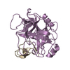
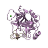
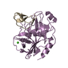
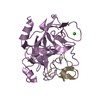
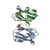
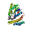
| Structure viewer | Molecule: MolmilJmol/JSmol |
|---|
- Downloads & links
-Download
| PDBx/mmCIF format | 1lu0.cif.gz | 43.1 KB | Display | PDBx/mmCIF format |
|---|---|---|---|---|
| PDB format | pdb1lu0.ent.gz | 30.3 KB | Display | PDB format |
| PDBx/mmJSON format | 1lu0.json.gz | Tree view | PDBx/mmJSON format | |
| Others |  Other downloads Other downloads |
-Validation report
| Arichive directory | https://data.pdbj.org/pub/pdb/validation_reports/lu/1lu0ftp://data.pdbj.org/pub/pdb/validation_reports/lu/1lu0 | HTTPS FTP |
|---|
-Related structure data
| Related structure data | |
|---|---|
| Similar structure data |


-Links
 PDBj
PDBj- Assembly
| Deposited unit | 
| |||||||||
|---|---|---|---|---|---|---|---|---|---|---|
| 1 | 
| |||||||||
| Unit cell |
| |||||||||
| Components on special symmetry positions |
| |||||||||
| Details | Four molecules of the inhibitor coordinate a single zinc cation in a tetrahedral fashion via their Glu19 side chains. Two of them (molecules A and B) constitute the contents of the asymmetric unit. The full coordination sphere is generated by the operation: -X, -Y, Z, of a two-fold axis. The zinc cation has a special position (occupancy 0.5) on this two-fold axis. |
-Components
-Protein/peptide , 1 types, 2 molecules AB
| #1: Protein/peptide | Mass: 3261.881 Da / Num. of mol.: 2 / Mutation: M8L Source method: isolated from a genetically manipulated source Source: (gene. exp.) Cucurbita maxima (winter squash) / Plasmid: pAED4 / Production host:  |
|---|
-Non-polymers , 5 types, 81 molecules 


| #2: Chemical | ChemComp-ZN /  Mass: 65.409 Da / Num. of mol.: 1 / Source method: obtained synthetically / Formula: Zn Mass: 65.409 Da / Num. of mol.: 1 / Source method: obtained synthetically / Formula: Zn | ||||||
|---|---|---|---|---|---|---|---|
| #3: Chemical |  Mass: 92.094 Da / Num. of mol.: 2 / Source method: obtained synthetically / Formula: C3H8O3 Mass: 92.094 Da / Num. of mol.: 2 / Source method: obtained synthetically / Formula: C3H8O3#4: Chemical | ChemComp-SO4 / |  Mass: 96.063 Da / Num. of mol.: 1 / Source method: obtained synthetically / Formula: SO4 Mass: 96.063 Da / Num. of mol.: 1 / Source method: obtained synthetically / Formula: SO4#5: Chemical | ChemComp-MRD / ( |  Mass: 118.174 Da / Num. of mol.: 1 / Source method: obtained synthetically / Formula: C6H14O2 / Comment: precipitant*YM Mass: 118.174 Da / Num. of mol.: 1 / Source method: obtained synthetically / Formula: C6H14O2 / Comment: precipitant*YM#6: Water | ChemComp-HOH / | Mass: 18.015 Da / Num. of mol.: 76 / Source method: isolated from a natural source / Formula: H2O |
-Details
| Has protein modification | Y |
|---|
-Experimental details
-Experiment
| Experiment | Method: X-RAY DIFFRACTION / Number of used crystals: 1 |
|---|
- Sample preparation
| Crystal | Density Matthews: 2.23 Å3/Da / Density % sol: 44.8 % | ||||||||||||||||||||||||||||||||||||||||||
|---|---|---|---|---|---|---|---|---|---|---|---|---|---|---|---|---|---|---|---|---|---|---|---|---|---|---|---|---|---|---|---|---|---|---|---|---|---|---|---|---|---|---|---|
| Crystal grow | Temperature: 293 K / Method: vapor diffusion, hanging drop / pH: 6.5 Details: PEG 8000, zinc sulfate, cacodylate, MPD, pH 6.5, VAPOR DIFFUSION, HANGING DROP, temperature 293.0K | ||||||||||||||||||||||||||||||||||||||||||
| Crystal grow | *PLUS | ||||||||||||||||||||||||||||||||||||||||||
| Components of the solutions | *PLUS
|
-Data collection
| Diffraction | Mean temperature: 100 K |
|---|---|
| Diffraction source | Source: SYNCHROTRON / Site: EMBL/DESY, HAMBURG  / Beamline: BW7A / Wavelength: 0.8919 Å / Beamline: BW7A / Wavelength: 0.8919 Å |
| Detector | Type: MARRESEARCH / Detector: CCD / Date: Apr 7, 2000 |
| Radiation | Monochromator: Si, double crystal, tunable / Protocol: SINGLE WAVELENGTH / Monochromatic (M) / Laue (L): M / Scattering type: x-ray |
| Radiation wavelength | Wavelength: 0.8919 Å / Relative weight: 1 |
| Reflection | Resolution: 1.03→15 Å / Num. all: 27045 / Num. obs: 27045 / % possible obs: 99.2 % / Observed criterion σ(F): 0 / Observed criterion σ(I): -3 / Redundancy: 9.3 % / Biso Wilson estimate: 12.9 Å2 / Rmerge(I) obs: 0.047 / Net I/σ(I): 30.7 |
| Reflection shell | Resolution: 1.03→1.07 Å / Redundancy: 3.5 % / Rmerge(I) obs: 0.151 / Mean I/σ(I) obs: 2.73 / Num. unique all: 2637 / % possible all: 98.3 |
| Reflection | *PLUS Lowest resolution: 15 Å / Num. measured all: 252201 |
| Reflection shell | *PLUS % possible obs: 98.3 % |
- Processing
| Software |
| |||||||||||||||||||||||||||||||||
|---|---|---|---|---|---|---|---|---|---|---|---|---|---|---|---|---|---|---|---|---|---|---|---|---|---|---|---|---|---|---|---|---|---|---|
| Refinement | Method to determine structure: Dual-space recycling / Resolution: 1.03→10 Å / Num. parameters: 5179 / Num. restraintsaints: 6000 / Isotropic thermal model: Anisotropic / Cross valid method: FREE R / σ(F): 0 / σ(I): -3 / StereochEM target val spec case: CSD / Stereochemistry target values: ENGH AND HUBER Details: Anisotropic refinement without stereochemical restraints on main chain atoms with low Beq (<15 A**2). In both molecules (A and B) there is a salt bridge between the C-terminus and the Arg1 side chain. The Arg5 side chain in molecule A is modeled in two conformations. The Val21 and Tyr27 side chains in both molecules are modeled in double conformation. His25 has different protonation and conformation in the two molecules. The zinc and sulfate ions lie on a common two fold axis. One MPD and two glycerol molecules are modeled in the solvent region. Blocked full-matix least-squares calculations were performed at the conclusion of the refinement. In remark 500, the atoms in all cases represent complementary pairs with partial occupancies.
| |||||||||||||||||||||||||||||||||
| Solvent computation | Solvent model: MOEWS & KRETSINGER, J.MOL.BIOL.91(1973)201-228 | |||||||||||||||||||||||||||||||||
| Refine analyze | Num. disordered residues: 5 / Occupancy sum hydrogen: 432 / Occupancy sum non hydrogen: 517.49 | |||||||||||||||||||||||||||||||||
| Refinement step | Cycle: LAST / Resolution: 1.03→10 Å
| |||||||||||||||||||||||||||||||||
| Refine LS restraints |
| |||||||||||||||||||||||||||||||||
| Software | *PLUS Name: SHELXL / Version: 97 / Classification: refinement | |||||||||||||||||||||||||||||||||
| Refinement | *PLUS Lowest resolution: 10 Å / Rfactor all: 0.12 / Rfactor Rfree: 0.1395 / Rfactor Rwork: 0.1141 | |||||||||||||||||||||||||||||||||
| Solvent computation | *PLUS | |||||||||||||||||||||||||||||||||
| Displacement parameters | *PLUS | |||||||||||||||||||||||||||||||||
| Refine LS restraints | *PLUS
|
