 Movie
Movie Controller

[English] 日本語
 Yorodumi
- PDB-1ls9: Structure of the Cytochrome c6 from the Green Alga Cladophora glo... -
+ Open data

- Basic information
| Entry | Database: PDB / ID: 1ls9 | ||||||
|---|---|---|---|---|---|---|---|
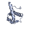
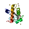
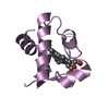
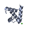
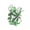
| Title | Structure of the Cytochrome c6 from the Green Alga Cladophora glomerata | ||||||
 Components Components | CYTOCHROME C6 | ||||||
 Keywords Keywords | ELECTRON TRANSPORT / omega loop / antiparallel beta-sheet / PROTOPORPHYRIN IX CONTAINING FE / heme / haem / CYTOCHROME | ||||||
| Function / homology |  Function and homology information Function and homology informationchloroplast thylakoid lumen / photosynthesis / electron transfer activity / iron ion binding / heme binding Similarity search - Function | ||||||
| Biological species |  Cladophora glomerata (plant) Cladophora glomerata (plant) | ||||||
| Method |  X-RAY DIFFRACTION / SYNCHROTRON / MOLECULAR REPLACEMENT / Resolution: 1.3 Å X-RAY DIFFRACTION / SYNCHROTRON / MOLECULAR REPLACEMENT / Resolution: 1.3 Å | ||||||
 Authors Authors | Carpentier, W. | ||||||
 Citation Citation | Journal: Biochemistry / Year: 2002 Title: Structural Basis for the Molecular Properties of Cytochrome C(6) Authors: Dikiy, A. / Carpentier, W. / Vandenberghe, I. / Borsari, M. / Safarov, N. / Dikaya, E. / Van Beeumen, J. / Ciurli, S. | ||||||
| History |
|
- Structure visualization
| Structure viewer | Molecule: MolmilJmol/JSmol |
|---|
- Downloads & links
-Download
| PDBx/mmCIF format | 1ls9.cif.gz | 58.4 KB | Display | PDBx/mmCIF format |
|---|---|---|---|---|
| PDB format | pdb1ls9.ent.gz | 41.6 KB | Display | PDB format |
| PDBx/mmJSON format | 1ls9.json.gz | Tree view | PDBx/mmJSON format | |
| Others |  Other downloads Other downloads |
-Validation report
| Arichive directory | https://data.pdbj.org/pub/pdb/validation_reports/ls/1ls9ftp://data.pdbj.org/pub/pdb/validation_reports/ls/1ls9 | HTTPS FTP |
|---|
-Related structure data
| Related structure data |  1ctjS S: Starting model for refinement |
|---|---|
| Similar structure data |


-Links
 PDBj
PDBj


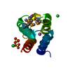
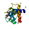
- Assembly
| Deposited unit | 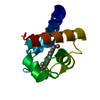
| ||||||||
|---|---|---|---|---|---|---|---|---|---|
| 1 |
| ||||||||
| Unit cell |
|
-Components
| #1: Protein | Mass: 9843.980 Da / Num. of mol.: 1 / Source method: isolated from a natural source / Source: (natural) Cladophora glomerata (plant) / References: UniProt: P83391 |
|---|---|
| #2: Chemical | ChemComp-HEM /   Mass: 616.487 Da / Num. of mol.: 1 / Source method: obtained synthetically / Formula: C34H32FeN4O4 Mass: 616.487 Da / Num. of mol.: 1 / Source method: obtained synthetically / Formula: C34H32FeN4O4 |
| #3: Water | ChemComp-HOH /  Mass: 18.015 Da / Num. of mol.: 200 / Source method: isolated from a natural source / Formula: H2O Mass: 18.015 Da / Num. of mol.: 200 / Source method: isolated from a natural source / Formula: H2O |
| Has protein modification | Y |
-Experimental details
-Experiment
| Experiment | Method: X-RAY DIFFRACTION / Number of used crystals: 1 |
|---|
- Sample preparation
| Crystal | Density Matthews: 2.08 Å3/Da / Density % sol: 40.31 % | ||||||||||||||||||||||||||||||||||||
|---|---|---|---|---|---|---|---|---|---|---|---|---|---|---|---|---|---|---|---|---|---|---|---|---|---|---|---|---|---|---|---|---|---|---|---|---|---|
| Crystal grow | Temperature: 277 K / Method: vapor diffusion, hanging drop / pH: 7.6 Details: 0.2 M Na-acetate, 0.1 M Na-cacodylate, pH 6.5, 30% w/v PEG 8000, pH 7.6, VAPOR DIFFUSION, HANGING DROP, temperature 277K | ||||||||||||||||||||||||||||||||||||
| Crystal grow | *PLUS Temperature: 4 ℃ | ||||||||||||||||||||||||||||||||||||
| Components of the solutions | *PLUS
|
-Data collection
| Diffraction | Mean temperature: 100 K |
|---|---|
| Diffraction source | Source: SYNCHROTRON / Site: EMBL/DESY, HAMBURG  / Beamline: X13 / Wavelength: 0.8015 Å / Beamline: X13 / Wavelength: 0.8015 Å |
| Detector | Type: MARRESEARCH / Detector: IMAGE PLATE / Date: May 17, 2001 / Details: 165-mm MAR CCD |
| Radiation | Monochromator: Triangular, Bent mirror / Protocol: SINGLE WAVELENGTH / Monochromatic (M) / Laue (L): M / Scattering type: x-ray |
| Radiation wavelength | Wavelength: 0.8015 Å / Relative weight: 1 |
| Reflection | Resolution: 1.3→14.8906 Å / Num. obs: 27067 / % possible obs: 98.4 % / Redundancy: 20.25 % / Rsym value: 0.053 / Net I/σ(I): 25.3 |
| Reflection shell | Resolution: 1.3→1.32 Å / Redundancy: 38.68 % / Mean I/σ(I) obs: 3.6 / Rsym value: 0.311 / % possible all: 97.6 |
| Reflection | *PLUS Lowest resolution: 14891 Å / Num. measured all: 114176 / Rmerge(I) obs: 0.053 |
| Reflection shell | *PLUS % possible obs: 97.6 % |
- Processing
| Software |
| ||||||||||||||||||||||||||||||||||||||||||||||||||||||||||||||||||||||||||||||||||||
|---|---|---|---|---|---|---|---|---|---|---|---|---|---|---|---|---|---|---|---|---|---|---|---|---|---|---|---|---|---|---|---|---|---|---|---|---|---|---|---|---|---|---|---|---|---|---|---|---|---|---|---|---|---|---|---|---|---|---|---|---|---|---|---|---|---|---|---|---|---|---|---|---|---|---|---|---|---|---|---|---|---|---|---|---|---|
| Refinement | Method to determine structure: MOLECULAR REPLACEMENT Starting model: PDB ENTRY 1CTJ Resolution: 1.3→14.684 Å / SU B: 0.55284 / SU ML: 0.02413 / Cross valid method: THROUGHOUT / ESU R: 0.0435 / ESU R Free: 0.04757 / Stereochemistry target values: Engh & Huber
| ||||||||||||||||||||||||||||||||||||||||||||||||||||||||||||||||||||||||||||||||||||
| Displacement parameters | Biso mean: 16.595 Å2
| ||||||||||||||||||||||||||||||||||||||||||||||||||||||||||||||||||||||||||||||||||||
| Refinement step | Cycle: LAST / Resolution: 1.3→14.684 Å
| ||||||||||||||||||||||||||||||||||||||||||||||||||||||||||||||||||||||||||||||||||||
| Refine LS restraints |
| ||||||||||||||||||||||||||||||||||||||||||||||||||||||||||||||||||||||||||||||||||||
| LS refinement shell | Resolution: 1.3→1.32 Å | ||||||||||||||||||||||||||||||||||||||||||||||||||||||||||||||||||||||||||||||||||||
| Refinement | *PLUS % reflection Rfree: 5 % / Rfactor Rfree: 0.19 / Rfactor Rwork: 0.143 | ||||||||||||||||||||||||||||||||||||||||||||||||||||||||||||||||||||||||||||||||||||
| Solvent computation | *PLUS | ||||||||||||||||||||||||||||||||||||||||||||||||||||||||||||||||||||||||||||||||||||
| Displacement parameters | *PLUS |
