 Movie
Movie Controller


+ Open data

- Basic information
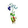
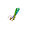
| Entry | Database: PDB / ID: 1lr9 | ||||||
|---|---|---|---|---|---|---|---|
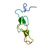
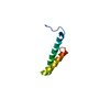
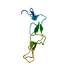
| Title | STRUCTURE OF Fs1, THE HEPARIN-BINDING DOMAIN OF FOLLISTATIN | ||||||
 Components Components | Follistatin | ||||||
 Keywords Keywords | HORMONE/GROWTH FACTOR / follistatin / heparin-binding / Fs1 / cystine-rich / HORMONE-GROWTH FACTOR COMPLEX | ||||||
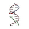
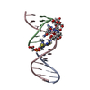
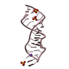
| Function / homology |  Function and homology information Function and homology informationAntagonism of Activin by Follistatin / activin receptor antagonist activity / negative regulation of follicle-stimulating hormone secretion / ameloblast differentiation / regulation of BMP signaling pathway / gamete generation / positive regulation of hair follicle development / activin binding / pattern specification process / negative regulation of activin receptor signaling pathway ...Antagonism of Activin by Follistatin / activin receptor antagonist activity / negative regulation of follicle-stimulating hormone secretion / ameloblast differentiation / regulation of BMP signaling pathway / gamete generation / positive regulation of hair follicle development / activin binding / pattern specification process / negative regulation of activin receptor signaling pathway / heparan sulfate proteoglycan binding / hair follicle morphogenesis / negative regulation of epithelial cell differentiation / odontogenesis of dentin-containing tooth / female gonad development / keratinocyte proliferation / BMP signaling pathway / hematopoietic progenitor cell differentiation / skeletal system development / cellular response to growth factor stimulus / cell differentiation / nucleolus / negative regulation of transcription by RNA polymerase II / : / extracellular region / nucleus / cytoplasm Similarity search - Function | ||||||
| Biological species |  | ||||||
| Method |  X-RAY DIFFRACTION / SYNCHROTRON / MOLECULAR REPLACEMENT / Resolution: 2.5 Å X-RAY DIFFRACTION / SYNCHROTRON / MOLECULAR REPLACEMENT / Resolution: 2.5 Å | ||||||
 Authors Authors | Innis, C.A. / Hyvonen, M. | ||||||
 Citation Citation | Journal: J.Biol.Chem. / Year: 2003 Title: Crystal Structures of the Heparan Sulfate-binding Domain of Follistatin: Insights into ligand binding. Authors: Innis, C.A. / Hyvonen, M. | ||||||
| History |
|
- Structure visualization
| Structure viewer | Molecule: MolmilJmol/JSmol |
|---|
- Downloads & links
-Download
| PDBx/mmCIF format | 1lr9.cif.gz | 26.2 KB | Display | PDBx/mmCIF format |
|---|---|---|---|---|
| PDB format | pdb1lr9.ent.gz | 16.4 KB | Display | PDB format |
| PDBx/mmJSON format | 1lr9.json.gz | Tree view | PDBx/mmJSON format | |
| Others |  Other downloads Other downloads |
-Validation report
| Arichive directory | https://data.pdbj.org/pub/pdb/validation_reports/lr/1lr9ftp://data.pdbj.org/pub/pdb/validation_reports/lr/1lr9 | HTTPS FTP |
|---|
-Related structure data
| Related structure data |  1lr7SC 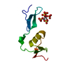 1lr8C S: Starting model for refinement C: citing same article ( |
|---|---|
| Similar structure data |

-Links
 PDBj
PDBj
- Assembly
| Deposited unit | 
| ||||||||
|---|---|---|---|---|---|---|---|---|---|
| 1 |
| ||||||||
| Unit cell |
|
-Components
| #1: Protein | Mass: 8342.812 Da / Num. of mol.: 1 / Fragment: Heparin binding domain Source method: isolated from a genetically manipulated source Source: (gene. exp.)  |
|---|---|
| #2: Water | ChemComp-HOH /  Mass: 18.015 Da / Num. of mol.: 28 / Source method: isolated from a natural source / Formula: H2O Mass: 18.015 Da / Num. of mol.: 28 / Source method: isolated from a natural source / Formula: H2O |
| Has protein modification | Y |
-Experimental details
-Experiment
| Experiment | Method: X-RAY DIFFRACTION / Number of used crystals: 1 |
|---|
- Sample preparation
| Crystal | Density Matthews: 2.35 Å3/Da / Density % sol: 43.68 % | |||||||||||||||||||||||||||||||||||
|---|---|---|---|---|---|---|---|---|---|---|---|---|---|---|---|---|---|---|---|---|---|---|---|---|---|---|---|---|---|---|---|---|---|---|---|---|
| Crystal grow | Temperature: 289 K / Method: vapor diffusion, hanging drop / pH: 8.5 Details: 30-35% PEG4000, 0.4 M Magnesium chloride, 0.1 M Tris-HCl, pH 8.5, VAPOR DIFFUSION, HANGING DROP, temperature 289K | |||||||||||||||||||||||||||||||||||
| Crystal grow | *PLUS Temperature: 18 ℃ / Method: vapor diffusion, hanging drop | |||||||||||||||||||||||||||||||||||
| Components of the solutions | *PLUS
|
-Data collection
| Diffraction | Mean temperature: 100 K |
|---|---|
| Diffraction source | Source: SYNCHROTRON / Site: ESRF  / Beamline: ID14-4 / Wavelength: 0.9795 Å / Beamline: ID14-4 / Wavelength: 0.9795 Å |
| Detector | Type: ADSC QUANTUM 4 / Detector: CCD / Date: Feb 11, 2001 / Details: Mirrors |
| Radiation | Monochromator: Si 111 CHANNEL / Protocol: SINGLE WAVELENGTH / Monochromatic (M) / Laue (L): M / Scattering type: x-ray |
| Radiation wavelength | Wavelength: 0.9795 Å / Relative weight: 1 |
| Reflection | Resolution: 2.5→38.6 Å / Num. obs: 2787 / % possible obs: 98.2 % / Observed criterion σ(F): 0 / Observed criterion σ(I): 0 / Redundancy: 7.8 % / Biso Wilson estimate: 33.26 Å2 / Rmerge(I) obs: 0.1 |
| Reflection shell | Resolution: 2.5→2.59 Å / Rmerge(I) obs: 0.171 / % possible all: 90.4 |
| Reflection | *PLUS Highest resolution: 2.5 Å / Lowest resolution: 38.6 Å / Rmerge(I) obs: 0.1 |
| Reflection shell | *PLUS Highest resolution: 2.5 Å / Lowest resolution: 2.59 Å / % possible obs: 90.4 % / Rmerge(I) obs: 0.17 |
- Processing
| Software |
| ||||||||||||||||||||||||||||||||||||||||||||||||||||||||||||||||||||||||||||||||||||||||||||||||||||||||||||||||||||||||||||||||||
|---|---|---|---|---|---|---|---|---|---|---|---|---|---|---|---|---|---|---|---|---|---|---|---|---|---|---|---|---|---|---|---|---|---|---|---|---|---|---|---|---|---|---|---|---|---|---|---|---|---|---|---|---|---|---|---|---|---|---|---|---|---|---|---|---|---|---|---|---|---|---|---|---|---|---|---|---|---|---|---|---|---|---|---|---|---|---|---|---|---|---|---|---|---|---|---|---|---|---|---|---|---|---|---|---|---|---|---|---|---|---|---|---|---|---|---|---|---|---|---|---|---|---|---|---|---|---|---|---|---|---|---|
| Refinement | Method to determine structure: MOLECULAR REPLACEMENT Starting model: PDB ENTRY 1LR7 Resolution: 2.5→38.63 Å / Cor.coef. Fo:Fc: 0.913 / Cor.coef. Fo:Fc free: 0.856 / SU B: 12.462 / SU ML: 0.287 / Isotropic thermal model: isotropic / Cross valid method: THROUGHOUT / σ(F): 0 / ESU R: 0.65 / ESU R Free: 0.302 / Stereochemistry target values: MAXIMUM LIKELIHOOD / Details: HYDROGENS HAVE BEEN ADDED IN THE RIDING POSITIONS
| ||||||||||||||||||||||||||||||||||||||||||||||||||||||||||||||||||||||||||||||||||||||||||||||||||||||||||||||||||||||||||||||||||
| Solvent computation | Ion probe radii: 0.8 Å / Shrinkage radii: 0.8 Å / VDW probe radii: 1.4 Å / Solvent model: BABINET MODEL WITH MASK | ||||||||||||||||||||||||||||||||||||||||||||||||||||||||||||||||||||||||||||||||||||||||||||||||||||||||||||||||||||||||||||||||||
| Displacement parameters | Biso mean: 24.605 Å2
| ||||||||||||||||||||||||||||||||||||||||||||||||||||||||||||||||||||||||||||||||||||||||||||||||||||||||||||||||||||||||||||||||||
| Refinement step | Cycle: LAST / Resolution: 2.5→38.63 Å
| ||||||||||||||||||||||||||||||||||||||||||||||||||||||||||||||||||||||||||||||||||||||||||||||||||||||||||||||||||||||||||||||||||
| Refine LS restraints |
| ||||||||||||||||||||||||||||||||||||||||||||||||||||||||||||||||||||||||||||||||||||||||||||||||||||||||||||||||||||||||||||||||||
| LS refinement shell | Resolution: 2.5→2.565 Å / Total num. of bins used: 20
| ||||||||||||||||||||||||||||||||||||||||||||||||||||||||||||||||||||||||||||||||||||||||||||||||||||||||||||||||||||||||||||||||||
| Refinement | *PLUS Highest resolution: 2.5 Å / Lowest resolution: 38.6 Å / Num. reflection obs: 2639 / Rfactor Rfree: 0.258 / Rfactor Rwork: 0.22 | ||||||||||||||||||||||||||||||||||||||||||||||||||||||||||||||||||||||||||||||||||||||||||||||||||||||||||||||||||||||||||||||||||
| Solvent computation | *PLUS | ||||||||||||||||||||||||||||||||||||||||||||||||||||||||||||||||||||||||||||||||||||||||||||||||||||||||||||||||||||||||||||||||||
| Displacement parameters | *PLUS | ||||||||||||||||||||||||||||||||||||||||||||||||||||||||||||||||||||||||||||||||||||||||||||||||||||||||||||||||||||||||||||||||||
| Refine LS restraints | *PLUS
|
