 Movie
Movie Controller

[English] 日本語
 Yorodumi
- PDB-1lok: The 1.20 Angstrom Resolution Crystal Structure of the Aminopeptid... -
+ Open data

- Basic information
| Entry | Database: PDB / ID: 1lok | ||||||
|---|---|---|---|---|---|---|---|
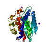
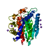
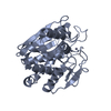
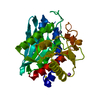
| Title | The 1.20 Angstrom Resolution Crystal Structure of the Aminopeptidase from Aeromonas proteolytica Complexed with Tris: A Tale of Buffer Inhibition | ||||||
 Components Components | Bacterial leucyl aminopeptidase | ||||||
 Keywords Keywords | HYDROLASE / metalloenzyme / aminopeptidase / tris / high resolution / metal coordination | ||||||
| Function / homology |  Function and homology information Function and homology informationbacterial leucyl aminopeptidase / metalloexopeptidase activity / aminopeptidase activity / proteolysis / extracellular region / metal ion binding Similarity search - Function | ||||||
| Biological species |  Vibrio proteolyticus (bacteria) Vibrio proteolyticus (bacteria) | ||||||
| Method |  X-RAY DIFFRACTION / SYNCHROTRON / FOURIER SYNTHESIS / Resolution: 1.2 Å X-RAY DIFFRACTION / SYNCHROTRON / FOURIER SYNTHESIS / Resolution: 1.2 Å | ||||||
 Authors Authors | Desmarais, W.T. / Bienvenue, D.L. / Bzymek, K.P. / Holz, R.C. / Petsko, G.A. / Ringe, D. | ||||||
 Citation Citation | Journal: Structure / Year: 2002 Title: The 1.20 Angstrom Resolution Crystal Structure of the Aminopeptidase from Aeromonas proteolytica Complexed with Tris A tale of Buffer Inhibition Authors: Desmarais, W.T. / Bienvenue, D.L. / Bzymek, K.P. / Holz, R.C. / Petsko, G.A. / Ringe, D. | ||||||
| History |
|
- Structure visualization
| Structure viewer | Molecule: MolmilJmol/JSmol |
|---|
- Downloads & links
-Download
| PDBx/mmCIF format | 1lok.cif.gz | 141.2 KB | Display | PDBx/mmCIF format |
|---|---|---|---|---|
| PDB format | pdb1lok.ent.gz | 110 KB | Display | PDB format |
| PDBx/mmJSON format | 1lok.json.gz | Tree view | PDBx/mmJSON format | |
| Others |  Other downloads Other downloads |
-Validation report
| Arichive directory | https://data.pdbj.org/pub/pdb/validation_reports/lo/1lokftp://data.pdbj.org/pub/pdb/validation_reports/lo/1lok | HTTPS FTP |
|---|
-Related structure data
| Related structure data |  1ampS S: Starting model for refinement |
|---|---|
| Similar structure data |


-Links
 PDBj
PDBj

- Assembly
| Deposited unit | 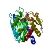
| ||||||||||||||||||
|---|---|---|---|---|---|---|---|---|---|---|---|---|---|---|---|---|---|---|---|
| 1 |
| ||||||||||||||||||
| Unit cell |
| ||||||||||||||||||
| Components on special symmetry positions |
| ||||||||||||||||||
| Details | The biological assembly is a monomer. |
-Components
-Protein , 1 types, 1 molecules A
| #1: Protein | Mass: 31427.350 Da / Num. of mol.: 1 / Source method: isolated from a natural source / Source: (natural) Vibrio proteolyticus (bacteria)References: UniProt: Q01693, bacterial leucyl aminopeptidase |
|---|
-Non-polymers , 5 types, 353 molecules 


| #2: Chemical |  Mass: 65.409 Da / Num. of mol.: 2 / Source method: obtained synthetically / Formula: Zn Mass: 65.409 Da / Num. of mol.: 2 / Source method: obtained synthetically / Formula: Zn#3: Chemical | ChemComp-NA / |  Mass: 22.990 Da / Num. of mol.: 1 / Source method: obtained synthetically / Formula: Na Mass: 22.990 Da / Num. of mol.: 1 / Source method: obtained synthetically / Formula: Na#4: Chemical | ChemComp-SCN / |  Mass: 58.082 Da / Num. of mol.: 1 / Source method: obtained synthetically / Formula: CNS Mass: 58.082 Da / Num. of mol.: 1 / Source method: obtained synthetically / Formula: CNS#5: Chemical | ChemComp-TRS / |  Mass: 122.143 Da / Num. of mol.: 1 / Source method: obtained synthetically / Formula: C4H12NO3 / Comment: pH buffer*YM Mass: 122.143 Da / Num. of mol.: 1 / Source method: obtained synthetically / Formula: C4H12NO3 / Comment: pH buffer*YM#6: Water | ChemComp-HOH / | Mass: 18.015 Da / Num. of mol.: 348 / Source method: isolated from a natural source / Formula: H2O |
|---|
-Details
| Has protein modification | Y |
|---|
-Experimental details
-Experiment
| Experiment | Method: X-RAY DIFFRACTION / Number of used crystals: 1 |
|---|
- Sample preparation
| Crystal | Density Matthews: 2.52 Å3/Da / Density % sol: 51.18 % | ||||||||||||||||||||||||||||||||||||||||||||||||||||||||
|---|---|---|---|---|---|---|---|---|---|---|---|---|---|---|---|---|---|---|---|---|---|---|---|---|---|---|---|---|---|---|---|---|---|---|---|---|---|---|---|---|---|---|---|---|---|---|---|---|---|---|---|---|---|---|---|---|---|
| Crystal grow | Temperature: 295 K / Method: vapor diffusion, hanging drop / pH: 8 Details: Tris, potassium thiocyanate, sodium chloride, pH 8.0, VAPOR DIFFUSION, HANGING DROP, temperature 295K | ||||||||||||||||||||||||||||||||||||||||||||||||||||||||
| Crystal grow | *PLUS Method: vapor diffusion | ||||||||||||||||||||||||||||||||||||||||||||||||||||||||
| Components of the solutions | *PLUS
|
-Data collection
| Diffraction | Mean temperature: 100 K |
|---|---|
| Diffraction source | Source: SYNCHROTRON / Site: APS  / Beamline: 14-BM-C / Wavelength: 1 Å / Beamline: 14-BM-C / Wavelength: 1 Å |
| Detector | Type: ADSC QUANTUM 4 / Detector: CCD / Date: Feb 11, 2000 |
| Radiation | Monochromator: Germanium / Protocol: SINGLE WAVELENGTH / Monochromatic (M) / Laue (L): M / Scattering type: x-ray |
| Radiation wavelength | Wavelength: 1 Å / Relative weight: 1 |
| Reflection | Resolution: 0.94→27.08 Å / Num. all: 89053 / Num. obs: 89053 / % possible obs: 88.4 % / Observed criterion σ(F): 0 / Observed criterion σ(I): 0 / Redundancy: 15 % |
| Reflection shell | Resolution: 0.94→1.25 Å / % possible all: 98 |
| Reflection | *PLUS Highest resolution: 1.2 Å / Lowest resolution: 30 Å / Num. obs: 117248 / % possible obs: 99 % / Num. measured all: 1348266 / Rmerge(I) obs: 0.057 |
| Reflection shell | *PLUS Highest resolution: 1.2 Å / % possible obs: 98 % / Rmerge(I) obs: 0.29 / Mean I/σ(I) obs: 3 |
- Processing
| Software |
| ||||||||||||||||||||
|---|---|---|---|---|---|---|---|---|---|---|---|---|---|---|---|---|---|---|---|---|---|
| Refinement | Method to determine structure: FOURIER SYNTHESIS Starting model: PDB ENTRY 1AMP Resolution: 1.2→27.08 Å / Isotropic thermal model: ANISOTROPIC / Cross valid method: THROUGHOUT / σ(F): 0 / σ(I): 0 / Stereochemistry target values: Engh & Huber
| ||||||||||||||||||||
| Refinement step | Cycle: LAST / Resolution: 1.2→27.08 Å
| ||||||||||||||||||||
| Refine LS restraints |
| ||||||||||||||||||||
| Software | *PLUS Name: SHELXL / Version: 97 / Classification: refinement | ||||||||||||||||||||
| Refinement | *PLUS Lowest resolution: 30 Å / Rfactor Rfree: 0.176 / Rfactor Rwork: 0.14 | ||||||||||||||||||||
| Solvent computation | *PLUS | ||||||||||||||||||||
| Displacement parameters | *PLUS |
