 ムービー
ムービー コントローラー


+ データを開く

- 基本情報
基本情報
| 登録情報 | データベース: PDB / ID: 1lf8 | ||||||
|---|---|---|---|---|---|---|---|
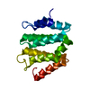
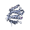
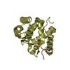
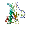
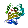
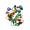
| タイトル | Complex of GGA3-VHS Domain and CI-MPR C-terminal Phosphopeptide | ||||||
 要素 要素 |
| ||||||
 キーワード キーワード | SIGNALING PROTEIN / VHS domain / Protein-phosphopeptide complex | ||||||
| 機能・相同性 |  機能・相同性情報 機能・相同性情報clathrin coat / positive regulation of lysosomal protein catabolic process / Retrograde transport at the Trans-Golgi-Network / response to tetrachloromethane / insulin-like growth factor receptor activity / retromer complex binding / insulin-like growth factor binding / protein transporter activity / insulin-like growth factor II binding / protein targeting to lysosome ...clathrin coat / positive regulation of lysosomal protein catabolic process / Retrograde transport at the Trans-Golgi-Network / response to tetrachloromethane / insulin-like growth factor receptor activity / retromer complex binding / insulin-like growth factor binding / protein transporter activity / insulin-like growth factor II binding / protein targeting to lysosome / trans-Golgi network transport vesicle / Golgi to plasma membrane transport / host-mediated activation of viral process / MET receptor recycling / retinoic acid binding / endocytic recycling / Golgi to plasma membrane protein transport / protein localization to cell surface / TBC/RABGAPs / lysosomal transport / Golgi Associated Vesicle Biogenesis / nuclear envelope lumen / D-mannose binding / negative regulation of amyloid-beta formation / endocytic vesicle / G-protein alpha-subunit binding / animal organ regeneration / response to retinoic acid / transport vesicle / phosphatidylinositol binding / receptor-mediated endocytosis / secretory granule membrane / ubiquitin binding / trans-Golgi network membrane / post-embryonic development / intracellular protein transport / protein catabolic process / phosphoprotein binding / trans-Golgi network / clathrin-coated endocytic vesicle membrane / liver development / protein destabilization / regulation of protein stability / small GTPase binding / recycling endosome membrane / late endosome / Cargo recognition for clathrin-mediated endocytosis / signaling receptor activity / Clathrin-mediated endocytosis / early endosome membrane / spermatogenesis / early endosome / lysosome / endosome / endosome membrane / positive regulation of apoptotic process / G protein-coupled receptor signaling pathway / Amyloid fiber formation / Golgi membrane / focal adhesion / Neutrophil degranulation / protein-containing complex binding / perinuclear region of cytoplasm / enzyme binding / cell surface / Golgi apparatus / signal transduction / protein-containing complex / extracellular exosome / identical protein binding / membrane / plasma membrane 類似検索 - 分子機能 | ||||||
| 生物種 |  Homo sapiens (ヒト) Homo sapiens (ヒト) | ||||||
| 手法 |  X線回折 / フーリエ合成 / 解像度: 2.3 Å X線回折 / フーリエ合成 / 解像度: 2.3 Å | ||||||
 データ登録者 データ登録者 | Kato, Y. / Misra, S. / Puertollano, R. / Hurley, J.H. / Bonifacino, J.S. | ||||||
 引用 引用 | ジャーナル: Nat.Struct.Biol. / 年: 2002 タイトル: Phosphoregulation of sorting signal-VHS domain interactions by a direct electrostatic mechanism. 著者: Kato, Y. / Misra, S. / Puertollano, R. / Hurley, J.H. / Bonifacino, J.S. | ||||||
| 履歴 |
|
- 構造の表示
構造の表示
| 構造ビューア | 分子: MolmilJmol/JSmol |
|---|
- ダウンロードとリンク
-ダウンロード
| PDBx/mmCIF形式 | 1lf8.cif.gz | 145 KB | 表示 | PDBx/mmCIF形式 |
|---|---|---|---|---|
| PDB形式 | pdb1lf8.ent.gz | 115.9 KB | 表示 | PDB形式 |
| PDBx/mmJSON形式 | 1lf8.json.gz | ツリー表示 | PDBx/mmJSON形式 | |
| その他 |  その他のダウンロード その他のダウンロード |
-検証レポート
| 文書・要旨 | 1lf8_validation.pdf.gz | 483 KB | 表示 | wwPDB検証レポート |
|---|---|---|---|---|
| 文書・詳細版 | 1lf8_full_validation.pdf.gz | 495.6 KB | 表示 | |
| XML形式データ | 1lf8_validation.xml.gz | 29.1 KB | 表示 | |
| CIF形式データ | 1lf8_validation.cif.gz | 41.1 KB | 表示 | |
| アーカイブディレクトリ | https://data.pdbj.org/pub/pdb/validation_reports/lf/1lf8ftp://data.pdbj.org/pub/pdb/validation_reports/lf/1lf8 | HTTPS FTP |
-関連構造データ
| 関連構造データ |  1juqS S: 精密化の開始モデル |
|---|---|
| 類似構造データ |


-リンク
 PDBj
PDBj


- 集合体
集合体
| 登録構造単位 | 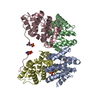
| ||||||||
|---|---|---|---|---|---|---|---|---|---|
| 1 | 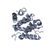
| ||||||||
| 2 | 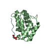
| ||||||||
| 3 | 
| ||||||||
| 4 | 
| ||||||||
| 単位格子 |
| ||||||||
| 詳細 | Each VHS domain/phosphopeptide complex corresponds to a single biological unit. There are the equivalent of 4 biological complexes per asymmetric unit. The apparent VHS domain dimer is likely a crystallization artifact. |
-要素
| #1: タンパク質 | 分子量: 19482.451 Da / 分子数: 4 / 断片: VHS domain (residues 1-166) / 由来タイプ: 組換発現 / 由来: (組換発現) Homo sapiens (ヒト) / プラスミド: pHis-parallel2 / 生物種 (発現宿主): Escherichia coli / 発現宿主:  #2: タンパク質・ペプチド | 分子量: 1537.478 Da / 分子数: 4 / 断片: C-terminus (residues 2480-2491) / 由来タイプ: 合成 詳細: The peptide was chemically synthesized. The sequence of the peptide is naturally found in Homo sapiens (Human). 参照: UniProt: P11717 #3: 水 | ChemComp-HOH / |  分子量: 18.015 Da / 分子数: 300 / 由来タイプ: 天然 / 式: H2O 分子量: 18.015 Da / 分子数: 300 / 由来タイプ: 天然 / 式: H2OHas protein modification | Y | |
|---|
-実験情報
-実験
| 実験 | 手法: X線回折 / 使用した結晶の数: 1 |
|---|
- 試料調製
試料調製
| 結晶 | マシュー密度: 2.61 Å3/Da / 溶媒含有率: 52.8 % | |||||||||||||||||||||||||||||||||||||||||||||||||
|---|---|---|---|---|---|---|---|---|---|---|---|---|---|---|---|---|---|---|---|---|---|---|---|---|---|---|---|---|---|---|---|---|---|---|---|---|---|---|---|---|---|---|---|---|---|---|---|---|---|---|
| 結晶化 | 温度: 293 K / 手法: 蒸気拡散法, ハンギングドロップ法 詳細: 100mM CAPS 10.2-11.0, 200 mM Lithium Sulfate, 1.3-2M Sodium/Potassium phosphate, VAPOR DIFFUSION, HANGING DROP, temperature 293K | |||||||||||||||||||||||||||||||||||||||||||||||||
| 結晶化 | *PLUS 温度: 20 ℃ / pH: 8 | |||||||||||||||||||||||||||||||||||||||||||||||||
| 溶液の組成 | *PLUS
|
-データ収集
| 回折 | 平均測定温度: 95 K |
|---|---|
| 放射光源 | 由来: 回転陽極 / タイプ: RIGAKU RU200 / 波長: 1.5418 Å |
| 検出器 | タイプ: RIGAKU RAXIS IV / 検出器: IMAGE PLATE / 日付: 2001年10月25日 / 詳細: Osmic Mirrors |
| 放射 | プロトコル: SINGLE WAVELENGTH / 単色(M)・ラウエ(L): M / 散乱光タイプ: x-ray |
| 放射波長 | 波長: 1.5418 Å / 相対比: 1 |
| 反射 | 解像度: 2.3→100 Å / Num. obs: 37720 / % possible obs: 96.1 % / Observed criterion σ(I): -3 / 冗長度: 6.1 % / Biso Wilson estimate: 31.5 Å2 / Rmerge(I) obs: 0.062 / Net I/σ(I): 20.5 |
| 反射 シェル | 解像度: 2.3→2.38 Å / Rmerge(I) obs: 0.356 / Mean I/σ(I) obs: 4.4 / Num. unique all: 3550 / % possible all: 91.5 |
| 反射 | *PLUS 最高解像度: 2.3 Å / 最低解像度: 50 Å / Rmerge(I) obs: 0.062 |
| 反射 シェル | *PLUS % possible obs: 91.5 % / Rmerge(I) obs: 0.356 |
- 解析
解析
| ソフトウェア |
| ||||||||||||||||||||||||||||||||||||
|---|---|---|---|---|---|---|---|---|---|---|---|---|---|---|---|---|---|---|---|---|---|---|---|---|---|---|---|---|---|---|---|---|---|---|---|---|---|
| 精密化 | 構造決定の手法: フーリエ合成 開始モデル: PDB ENTRY 1JUQ 解像度: 2.3→89.83 Å / Rfactor Rfree error: 0.004 / Data cutoff high absF: 713707.58 / Data cutoff low absF: 0 / Isotropic thermal model: RESTRAINED / 交差検証法: THROUGHOUT / σ(F): 0 / 立体化学のターゲット値: Engh & Huber
| ||||||||||||||||||||||||||||||||||||
| 溶媒の処理 | 溶媒モデル: FLAT MODEL / Bsol: 42.6607 Å2 / ksol: 0.338167 e/Å3 | ||||||||||||||||||||||||||||||||||||
| 原子変位パラメータ | Biso mean: 49.8 Å2
| ||||||||||||||||||||||||||||||||||||
| Refine analyze |
| ||||||||||||||||||||||||||||||||||||
| 精密化ステップ | サイクル: LAST / 解像度: 2.3→89.83 Å
| ||||||||||||||||||||||||||||||||||||
| 拘束条件 |
| ||||||||||||||||||||||||||||||||||||
| LS精密化 シェル | 解像度: 2.3→2.44 Å / Rfactor Rfree error: 0.014 / Total num. of bins used: 6
| ||||||||||||||||||||||||||||||||||||
| Xplor file |
| ||||||||||||||||||||||||||||||||||||
| 精密化 | *PLUS 最高解像度: 2.3 Å / 最低解像度: 50 Å / % reflection Rfree: 10 % / Rfactor Rfree: 0.255 / Rfactor Rwork: 0.211 | ||||||||||||||||||||||||||||||||||||
| 溶媒の処理 | *PLUS | ||||||||||||||||||||||||||||||||||||
| 原子変位パラメータ | *PLUS | ||||||||||||||||||||||||||||||||||||
| 拘束条件 | *PLUS
| ||||||||||||||||||||||||||||||||||||
| LS精密化 シェル | *PLUS Rfactor Rfree: 0.322 / Rfactor Rwork: 0.264 |
