 Movie
Movie Controller

[English] 日本語
 Yorodumi
- PDB-1lep: THREE-DIMENSIONAL STRUCTURE OF THE IMMUNODOMINANT HEAT-SHOCK PROT... -
+ Open data

- Basic information
| Entry | Database: PDB / ID: 1lep | ||||||
|---|---|---|---|---|---|---|---|
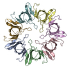
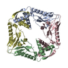
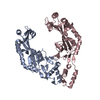
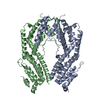
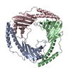
| Title | THREE-DIMENSIONAL STRUCTURE OF THE IMMUNODOMINANT HEAT-SHOCK PROTEIN CHAPERONIN-10 OF MYCOBACTERIUM LEPRAE | ||||||
 Components Components | CHAPERONIN-10 | ||||||
 Keywords Keywords | CHAPERONE / ANTIGEN / HEAT SHOCK | ||||||
| Function / homology |  Function and homology information Function and homology information: / protein folding chaperone / unfolded protein binding / protein-folding chaperone binding / ATP binding / metal ion binding / cytoplasm Similarity search - Function | ||||||
| Biological species |  Mycobacterium leprae (bacteria) Mycobacterium leprae (bacteria) | ||||||
| Method |  X-RAY DIFFRACTION / Resolution: 3.5 Å X-RAY DIFFRACTION / Resolution: 3.5 Å | ||||||
 Authors Authors | Mande, S.C. / Hol, W.G.J. | ||||||
 Citation Citation | Journal: Science / Year: 1996 Title: Structure of the heat shock protein chaperonin-10 of Mycobacterium leprae. Authors: Mande, S.C. / Mehra, V. / Bloom, B.R. / Hol, W.G. | ||||||
| History |
|
- Structure visualization
| Structure viewer | Molecule: MolmilJmol/JSmol |
|---|
- Downloads & links
-Download
| PDBx/mmCIF format | 1lep.cif.gz | 27.8 KB | Display | PDBx/mmCIF format |
|---|---|---|---|---|
| PDB format | pdb1lep.ent.gz | 16.4 KB | Display | PDB format |
| PDBx/mmJSON format | 1lep.json.gz | Tree view | PDBx/mmJSON format | |
| Others |  Other downloads Other downloads |
-Validation report
| Arichive directory | https://data.pdbj.org/pub/pdb/validation_reports/le/1lepftp://data.pdbj.org/pub/pdb/validation_reports/le/1lep | HTTPS FTP |
|---|
-Related structure data
| Similar structure data |
|---|


-Links
 PDBj
PDBj

- Assembly
| Deposited unit | 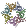
| ||||||||||||||||||||||||||||
|---|---|---|---|---|---|---|---|---|---|---|---|---|---|---|---|---|---|---|---|---|---|---|---|---|---|---|---|---|---|
| 1 |
| ||||||||||||||||||||||||||||
| Unit cell |
| ||||||||||||||||||||||||||||
| Noncrystallographic symmetry (NCS) | NCS oper:
|
-Components
| #1: Protein | Mass: 10682.150 Da / Num. of mol.: 7 / Source method: isolated from a natural source / Source: (natural) Mycobacterium leprae (bacteria) / References: UniProt: P24301 |
|---|
-Experimental details
-Experiment
| Experiment | Method: X-RAY DIFFRACTION |
|---|
- Sample preparation
| Crystal | Density Matthews: 2.66 Å3/Da / Density % sol: 55 % | ||||||||||||||||||||||||||||||||||||||||||
|---|---|---|---|---|---|---|---|---|---|---|---|---|---|---|---|---|---|---|---|---|---|---|---|---|---|---|---|---|---|---|---|---|---|---|---|---|---|---|---|---|---|---|---|
| Crystal | *PLUS | ||||||||||||||||||||||||||||||||||||||||||
| Crystal grow | *PLUS Temperature: 4 ℃ / pH: 6 / Method: vapor diffusion, hanging drop | ||||||||||||||||||||||||||||||||||||||||||
| Components of the solutions | *PLUS
|
-Data collection
| Diffraction source | Wavelength: 1.5418 |
|---|---|
| Detector | Type: RIGAKU RAXIS II / Detector: IMAGE PLATE / Date: 1995 |
| Radiation | Monochromatic (M) / Laue (L): M / Scattering type: x-ray |
| Radiation wavelength | Wavelength: 1.5418 Å / Relative weight: 1 |
| Reflection | Rmerge(I) obs: 0.088 |
| Reflection | *PLUS Highest resolution: 3.5 Å / % possible obs: 98 % |
| Reflection shell | *PLUS Highest resolution: 3.5 Å / Lowest resolution: 3.6 Å / % possible obs: 90 % |
- Processing
| Software |
| ||||||||||||||||||||||||||||||||||||||||||||||||||||||||||||
|---|---|---|---|---|---|---|---|---|---|---|---|---|---|---|---|---|---|---|---|---|---|---|---|---|---|---|---|---|---|---|---|---|---|---|---|---|---|---|---|---|---|---|---|---|---|---|---|---|---|---|---|---|---|---|---|---|---|---|---|---|---|
| Refinement | Resolution: 3.5→8 Å / σ(F): 2
| ||||||||||||||||||||||||||||||||||||||||||||||||||||||||||||
| Displacement parameters | Biso mean: 40 Å2 | ||||||||||||||||||||||||||||||||||||||||||||||||||||||||||||
| Refinement step | Cycle: LAST / Resolution: 3.5→8 Å
| ||||||||||||||||||||||||||||||||||||||||||||||||||||||||||||
| Refine LS restraints |
|
