 Movie
Movie Controller


+ Open data

- Basic information
| Entry | Database: PDB / ID: 1l7v | ||||||
|---|---|---|---|---|---|---|---|
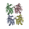
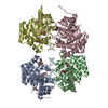
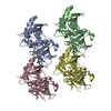
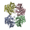
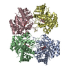
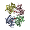
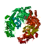
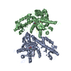
| Title | Bacterial ABC Transporter Involved in B12 Uptake | ||||||
 Components Components |
| ||||||
 Keywords Keywords | TRANSPORT PROTEIN/HYDROLASE / ABC transporter / integral membrane protein / ATP binding cassette / ATP hydrolysis / Vitamin B12 / TRANSPORT PROTEIN-HYDROLASE COMPLEX | ||||||
| Function / homology |  Function and homology information Function and homology informationBtuCD complex / ABC-type vitamin B12 transporter / cobalamin transport complex / ABC-type vitamin B12 transporter activity / cobalamin transport / extrinsic component of membrane / ATPase-coupled transmembrane transporter activity / transmembrane transporter activity / ATP-binding cassette (ABC) transporter complex / ATP hydrolysis activity ...BtuCD complex / ABC-type vitamin B12 transporter / cobalamin transport complex / ABC-type vitamin B12 transporter activity / cobalamin transport / extrinsic component of membrane / ATPase-coupled transmembrane transporter activity / transmembrane transporter activity / ATP-binding cassette (ABC) transporter complex / ATP hydrolysis activity / ATP binding / membrane / identical protein binding / plasma membrane Similarity search - Function | ||||||
| Biological species |  | ||||||
| Method |  X-RAY DIFFRACTION / SYNCHROTRON / MAD / Resolution: 3.2 Å X-RAY DIFFRACTION / SYNCHROTRON / MAD / Resolution: 3.2 Å | ||||||
 Authors Authors | Locher, K.P. / Lee, A.T. / Rees, D.C. | ||||||
 Citation Citation | Journal: Science / Year: 2002 Title: The E. coli BtuCD structure: a framework for ABC transporter architecture and mechanism. Authors: Locher, K.P. / Lee, A.T. / Rees, D.C. | ||||||
| History |
|
- Structure visualization
| Structure viewer | Molecule: MolmilJmol/JSmol |
|---|
- Downloads & links
-Download
| PDBx/mmCIF format | 1l7v.cif.gz | 219.9 KB | Display | PDBx/mmCIF format |
|---|---|---|---|---|
| PDB format | pdb1l7v.ent.gz | 176.3 KB | Display | PDB format |
| PDBx/mmJSON format | 1l7v.json.gz | Tree view | PDBx/mmJSON format | |
| Others |  Other downloads Other downloads |
-Validation report
| Arichive directory | https://data.pdbj.org/pub/pdb/validation_reports/l7/1l7vftp://data.pdbj.org/pub/pdb/validation_reports/l7/1l7v | HTTPS FTP |
|---|
-Related structure data
| Similar structure data |
|---|


-Links
 PDBj
PDBj

- Assembly
| Deposited unit | 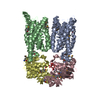
| ||||||||
|---|---|---|---|---|---|---|---|---|---|
| 1 |
| ||||||||
| Unit cell |
| ||||||||
| Details | Asymmetric unit contains full transporter: two copies each of BtuC and BtuD |
-Components
| #1: Protein | Mass: 35487.941 Da / Num. of mol.: 2 Source method: isolated from a genetically manipulated source Source: (gene. exp.) #2: Protein | Mass: 27536.234 Da / Num. of mol.: 2 Source method: isolated from a genetically manipulated source Source: (gene. exp.) #3: Chemical |   Mass: 395.759 Da / Num. of mol.: 2 / Source method: obtained synthetically / Formula: O12V4 Mass: 395.759 Da / Num. of mol.: 2 / Source method: obtained synthetically / Formula: O12V4Has protein modification | Y | |
|---|
-Experimental details
-Experiment
| Experiment | Method: X-RAY DIFFRACTION / Number of used crystals: 1 |
|---|
- Sample preparation
| Crystal | Density Matthews: 3.7 Å3/Da / Density % sol: 66.72 % | ||||||||||||||||||||||||||||||||||||
|---|---|---|---|---|---|---|---|---|---|---|---|---|---|---|---|---|---|---|---|---|---|---|---|---|---|---|---|---|---|---|---|---|---|---|---|---|---|
| Crystal grow | Temperature: 278 K / Method: vapor diffusion, sitting drop / pH: 8 Details: PEG 2000, LDAO, Tris, magnesium nitrate, MPD, D2O, pH 8.0, VAPOR DIFFUSION, SITTING DROP, temperature 278K | ||||||||||||||||||||||||||||||||||||
| Crystal grow | *PLUS | ||||||||||||||||||||||||||||||||||||
| Components of the solutions | *PLUS
|
-Data collection
| Diffraction | Mean temperature: 100 K |
|---|---|
| Diffraction source | Source: SYNCHROTRON / Site: SSRL  / Beamline: BL9-2 / Wavelength: 1.7712 Å / Beamline: BL9-2 / Wavelength: 1.7712 Å |
| Detector | Type: ADSC QUANTUM 4 / Detector: CCD / Date: Dec 18, 2001 |
| Radiation | Monochromator: SAGITALLY FOCUSED Si(111) / Protocol: SINGLE WAVELENGTH / Monochromatic (M) / Laue (L): M / Scattering type: x-ray |
| Radiation wavelength | Wavelength: 1.7712 Å / Relative weight: 1 |
| Reflection | Resolution: 3.2→30 Å / Num. all: 30814 / Num. obs: 30660 / % possible obs: 99.5 % / Observed criterion σ(F): 0 / Observed criterion σ(I): -3 |
| Reflection shell | Resolution: 3.2→3.29 Å / % possible all: 94.3 |
| Reflection | *PLUS Lowest resolution: 30 Å / Redundancy: 8.2 % / Rmerge(I) obs: 0.067 |
| Reflection shell | *PLUS % possible obs: 94.3 % / Rmerge(I) obs: 0.448 / Mean I/σ(I) obs: 4.1 |
- Processing
| Software |
| ||||||||||||||||||||
|---|---|---|---|---|---|---|---|---|---|---|---|---|---|---|---|---|---|---|---|---|---|
| Refinement | Method to determine structure: MAD / Resolution: 3.2→15 Å / σ(F): 0 / Stereochemistry target values: Engh & Huber
| ||||||||||||||||||||
| Refinement step | Cycle: LAST / Resolution: 3.2→15 Å
| ||||||||||||||||||||
| Refinement | *PLUS Lowest resolution: 15 Å / Rfactor obs: 0.262 / Rfactor Rfree: 0.286 / Rfactor Rwork: 0.262 | ||||||||||||||||||||
| Solvent computation | *PLUS | ||||||||||||||||||||
| Displacement parameters | *PLUS | ||||||||||||||||||||
| Refine LS restraints | *PLUS
|
