 Movie
Movie Controller

[English] 日本語
 Yorodumi
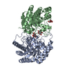
- PDB-2qum: Crystal structure of D-tagatose 3-epimerase from Pseudomonas cich... -
+ Open data

- Basic information
| Entry | Database: PDB / ID: 2qum | ||||||
|---|---|---|---|---|---|---|---|
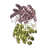
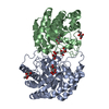
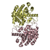
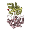
| Title | Crystal structure of D-tagatose 3-epimerase from Pseudomonas cichorii with D-tagatose | ||||||
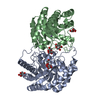
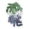
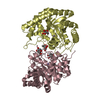
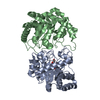
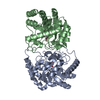
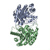
 Components Components | D-tagatose 3-epimerase | ||||||
 Keywords Keywords | ISOMERASE / BETA/ALPHA BARREL | ||||||
| Function / homology |  Function and homology information Function and homology information | ||||||
| Biological species |  Pseudomonas cichorii (bacteria) Pseudomonas cichorii (bacteria) | ||||||
| Method |  X-RAY DIFFRACTION / SYNCHROTRON / MOLECULAR REPLACEMENT / Resolution: 2.28 Å X-RAY DIFFRACTION / SYNCHROTRON / MOLECULAR REPLACEMENT / Resolution: 2.28 Å | ||||||
 Authors Authors | Yoshida, H. / Yamada, M. / Nishitani, T. / Takada, G. / Izumori, K. / Kamitori, S. | ||||||
 Citation Citation | Journal: J.Mol.Biol. / Year: 2007 Title: Crystal structures of D-tagatose 3-epimerase from Pseudomonas cichorii and its complexes with D-tagatose and D-fructose Authors: Yoshida, H. / Yamada, M. / Nishitani, T. / Takada, G. / Izumori, K. / Kamitori, S. | ||||||
| History |
|
- Structure visualization
| Structure viewer | Molecule: MolmilJmol/JSmol |
|---|
- Downloads & links
-Download
| PDBx/mmCIF format | 2qum.cif.gz | 249.4 KB | Display | PDBx/mmCIF format |
|---|---|---|---|---|
| PDB format | pdb2qum.ent.gz | 199.1 KB | Display | PDB format |
| PDBx/mmJSON format | 2qum.json.gz | Tree view | PDBx/mmJSON format | |
| Others |  Other downloads Other downloads |
-Validation report
| Arichive directory | https://data.pdbj.org/pub/pdb/validation_reports/qu/2qumftp://data.pdbj.org/pub/pdb/validation_reports/qu/2qum | HTTPS FTP |
|---|
-Related structure data
| Related structure data | 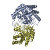 2ou4SC 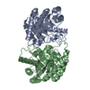 2qulC  2qunC C: citing same article ( S: Starting model for refinement |
|---|---|
| Similar structure data |


-Links
 PDBj
PDBj
- Assembly
| Deposited unit | 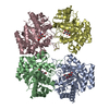
| ||||||||
|---|---|---|---|---|---|---|---|---|---|
| 1 | 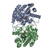
| ||||||||
| 2 | 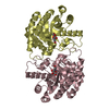
| ||||||||
| Unit cell |
|
-Components
| #1: Protein | Mass: 32657.404 Da / Num. of mol.: 4 Source method: isolated from a genetically manipulated source Source: (gene. exp.) Pseudomonas cichorii (bacteria) / Strain: ST-24 / Plasmid: pTrc99A / Production host: References: UniProt: O50580, Isomerases; Intramolecular oxidoreductases; Interconverting aldoses and ketoses, and related compounds #2: Sugar | ChemComp-TAG / 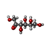  Type: D-saccharide / Mass: 180.156 Da / Num. of mol.: 4 Type: D-saccharide / Mass: 180.156 Da / Num. of mol.: 4Source method: isolated from a genetically manipulated source Formula: C6H12O6 #3: Chemical | ChemComp-MN /   Mass: 54.938 Da / Num. of mol.: 4 / Source method: obtained synthetically / Formula: Mn Mass: 54.938 Da / Num. of mol.: 4 / Source method: obtained synthetically / Formula: Mn#4: Water | ChemComp-HOH / |  Mass: 18.015 Da / Num. of mol.: 762 / Source method: isolated from a natural source / Formula: H2O Mass: 18.015 Da / Num. of mol.: 762 / Source method: isolated from a natural source / Formula: H2O |
|---|
-Experimental details
-Experiment
| Experiment | Method: X-RAY DIFFRACTION / Number of used crystals: 1 |
|---|
- Sample preparation
| Crystal | Density Matthews: 2.38 Å3/Da / Density % sol: 48.23 % |
|---|---|
| Crystal grow | Temperature: 298 K / Method: vapor diffusion, hanging drop / pH: 7.5 Details: 20% PEG 10000, 0.1M HEPES, 0.1M tri-sodium citrate, pH 7.5, VAPOR DIFFUSION, HANGING DROP, temperature 298.0K |
-Data collection
| Diffraction | Mean temperature: 100 K |
|---|---|
| Diffraction source | Source: SYNCHROTRON / Site: Photon Factory  / Beamline: BL-6A / Wavelength: 0.978 Å / Beamline: BL-6A / Wavelength: 0.978 Å |
| Detector | Type: ADSC QUANTUM 4 / Detector: CCD / Date: Jan 28, 2007 |
| Radiation | Monochromator: GRAPHITE / Protocol: SINGLE WAVELENGTH / Monochromatic (M) / Laue (L): M / Scattering type: x-ray |
| Radiation wavelength | Wavelength: 0.978 Å / Relative weight: 1 |
| Reflection | Resolution: 2.28→50 Å / Num. all: 55980 / Num. obs: 55980 / % possible obs: 99.4 % / Observed criterion σ(F): 0 / Observed criterion σ(I): 0 / Redundancy: 3.6 % / Biso Wilson estimate: 14.5 Å2 / Rmerge(I) obs: 0.099 / Net I/σ(I): 7.4 |
| Reflection shell | Resolution: 2.28→2.36 Å / Redundancy: 3.4 % / Rmerge(I) obs: 0.29 / Mean I/σ(I) obs: 4.3 / Num. unique all: 5571 / % possible all: 98.5 |
- Processing
| Software |
| |||||||||||||||||||||||||
|---|---|---|---|---|---|---|---|---|---|---|---|---|---|---|---|---|---|---|---|---|---|---|---|---|---|---|
| Refinement | Method to determine structure: MOLECULAR REPLACEMENT Starting model: PDB ENTRY 2OU4 Resolution: 2.28→41.31 Å / Rfactor Rfree error: 0.003 / Data cutoff high absF: 37837.84 / Data cutoff low absF: 0 / Isotropic thermal model: RESTRAINED / Cross valid method: THROUGHOUT / σ(F): 0 / Stereochemistry target values: Engh & Huber
| |||||||||||||||||||||||||
| Solvent computation | Solvent model: FLAT MODEL / Bsol: 56.0416 Å2 / ksol: 0.404938 e/Å3 | |||||||||||||||||||||||||
| Displacement parameters | Biso mean: 20.2 Å2
| |||||||||||||||||||||||||
| Refine analyze |
| |||||||||||||||||||||||||
| Refinement step | Cycle: LAST / Resolution: 2.28→41.31 Å
| |||||||||||||||||||||||||
| Refine LS restraints |
| |||||||||||||||||||||||||
| LS refinement shell | Resolution: 2.28→2.42 Å / Rfactor Rfree error: 0.01 / Total num. of bins used: 6
| |||||||||||||||||||||||||
| Xplor file |
|
