 Movie
Movie Controller

[English] 日本語
 Yorodumi
- PDB-5j8l: Crystal structure of D-tagatose 3-epimerase C66S from Pseudomonas... -
+ Open data

- Basic information
| Entry | Database: PDB / ID: 5j8l | |||||||||
|---|---|---|---|---|---|---|---|---|---|---|
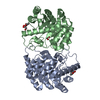
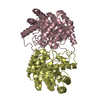
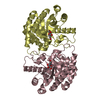
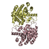
| Title | Crystal structure of D-tagatose 3-epimerase C66S from Pseudomonas cichorii in complex with 1-deoxy L-tagatose, using a crystal grown in microgravity | |||||||||
 Components Components | D-tagatose 3-epimerase | |||||||||
 Keywords Keywords | ISOMERASE / Epimerase | |||||||||
| Function / homology |  Function and homology information Function and homology information | |||||||||
| Biological species |  Pseudomonas cichorii (bacteria) Pseudomonas cichorii (bacteria) | |||||||||
| Method |  X-RAY DIFFRACTION / SYNCHROTRON / MOLECULAR REPLACEMENT / Resolution: 1.73 Å X-RAY DIFFRACTION / SYNCHROTRON / MOLECULAR REPLACEMENT / Resolution: 1.73 Å | |||||||||
 Authors Authors | Yoshida, H. / Yoshihara, A. / Izumori, K. / Kamitori, S. | |||||||||
 Citation Citation | Journal: Appl. Microbiol. Biotechnol. / Year: 2016 Title: X-ray structures of the Pseudomonas cichorii D-tagatose 3-epimerase mutant form C66S recognizing deoxy sugars as substrates Authors: Yoshida, H. / Yoshihara, A. / Ishii, T. / Izumori, K. / Kamitori, S. #1: Journal: J.Mol.Biol. / Year: 2007Title: Crystal structures of D-tagatose 3-epimerase from Pseudomonas cichorii and its complexes with D-tagatose and D-fructose. Authors: Yoshida, H. / Yamada, M. / Nishitani, T. / Takada, G. / Izumori, K. / Kamitori, S. | |||||||||
| History |
|
- Structure visualization
| Structure viewer | Molecule: MolmilJmol/JSmol |
|---|
- Downloads & links
-Download
| PDBx/mmCIF format | 5j8l.cif.gz | 257.8 KB | Display | PDBx/mmCIF format |
|---|---|---|---|---|
| PDB format | pdb5j8l.ent.gz | 205.7 KB | Display | PDB format |
| PDBx/mmJSON format | 5j8l.json.gz | Tree view | PDBx/mmJSON format | |
| Others |  Other downloads Other downloads |
-Validation report
| Arichive directory | https://data.pdbj.org/pub/pdb/validation_reports/j8/5j8lftp://data.pdbj.org/pub/pdb/validation_reports/j8/5j8l | HTTPS FTP |
|---|
-Related structure data
| Related structure data | 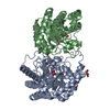 4xslC 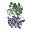 4xsmC 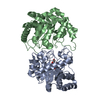 4ytqC 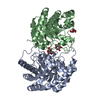 4ytrC  4ytsC  4yttC  4ytuC  2qulS C: citing same article ( S: Starting model for refinement |
|---|---|
| Similar structure data |


-Links
 PDBj
PDBj
- Assembly
| Deposited unit | 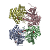
| ||||||||
|---|---|---|---|---|---|---|---|---|---|
| 1 | 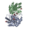
| ||||||||
| 2 | 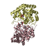
| ||||||||
| Unit cell |
|
-Components
| #1: Protein | Mass: 33858.613 Da / Num. of mol.: 4 / Mutation: C66S Source method: isolated from a genetically manipulated source Source: (gene. exp.) Pseudomonas cichorii (bacteria) / Plasmid: pQE60 / Production host: #2: Chemical | ChemComp-MN /   Mass: 54.938 Da / Num. of mol.: 4 / Source method: obtained synthetically / Formula: Mn Mass: 54.938 Da / Num. of mol.: 4 / Source method: obtained synthetically / Formula: Mn#3: Chemical |   Mass: 164.156 Da / Num. of mol.: 2 / Source method: obtained synthetically / Formula: C6H12O5 Mass: 164.156 Da / Num. of mol.: 2 / Source method: obtained synthetically / Formula: C6H12O5#4: Sugar | ChemComp-TGK /   Type: L-saccharide, beta linking / Mass: 164.156 Da / Num. of mol.: 8 Type: L-saccharide, beta linking / Mass: 164.156 Da / Num. of mol.: 8Source method: isolated from a genetically manipulated source Formula: C6H12O5 #5: Water | ChemComp-HOH / |  Mass: 18.015 Da / Num. of mol.: 921 / Source method: isolated from a natural source / Formula: H2O Mass: 18.015 Da / Num. of mol.: 921 / Source method: isolated from a natural source / Formula: H2O |
|---|
-Experimental details
-Experiment
| Experiment | Method: X-RAY DIFFRACTION / Number of used crystals: 1 |
|---|
- Sample preparation
| Crystal | Density Matthews: 2.41 Å3/Da / Density % sol: 48.97 % |
|---|---|
| Crystal grow | Temperature: 293 K / Method: liquid diffusion / pH: 4.6 / Details: 15 % PEG4000, 0.1 M Sodium acetate |
-Data collection
| Diffraction | Mean temperature: 100 K | ||||||||||||||||||||||||||||||||||||||||||||||||||||||||||||||||||||||||||||||||||||||||||||||||||||||||||||||||||||||||||||||
|---|---|---|---|---|---|---|---|---|---|---|---|---|---|---|---|---|---|---|---|---|---|---|---|---|---|---|---|---|---|---|---|---|---|---|---|---|---|---|---|---|---|---|---|---|---|---|---|---|---|---|---|---|---|---|---|---|---|---|---|---|---|---|---|---|---|---|---|---|---|---|---|---|---|---|---|---|---|---|---|---|---|---|---|---|---|---|---|---|---|---|---|---|---|---|---|---|---|---|---|---|---|---|---|---|---|---|---|---|---|---|---|---|---|---|---|---|---|---|---|---|---|---|---|---|---|---|---|
| Diffraction source | Source: SYNCHROTRON / Site: Photon Factory  / Beamline: BL-5A / Wavelength: 1 Å / Beamline: BL-5A / Wavelength: 1 Å | ||||||||||||||||||||||||||||||||||||||||||||||||||||||||||||||||||||||||||||||||||||||||||||||||||||||||||||||||||||||||||||||
| Detector | Type: ADSC QUANTUM 315 / Detector: CCD / Date: Dec 9, 2014 | ||||||||||||||||||||||||||||||||||||||||||||||||||||||||||||||||||||||||||||||||||||||||||||||||||||||||||||||||||||||||||||||
| Radiation | Protocol: SINGLE WAVELENGTH / Monochromatic (M) / Laue (L): M / Scattering type: x-ray | ||||||||||||||||||||||||||||||||||||||||||||||||||||||||||||||||||||||||||||||||||||||||||||||||||||||||||||||||||||||||||||||
| Radiation wavelength | Wavelength: 1 Å / Relative weight: 1 | ||||||||||||||||||||||||||||||||||||||||||||||||||||||||||||||||||||||||||||||||||||||||||||||||||||||||||||||||||||||||||||||
| Reflection | Resolution: 1.73→100 Å / Num. obs: 121443 / % possible obs: 92.1 % / Redundancy: 2 % / Biso Wilson estimate: 16.8 Å2 / Rmerge(I) obs: 0.075 / Net I/av σ(I): 15.773 / Net I/σ(I): 10.2 | ||||||||||||||||||||||||||||||||||||||||||||||||||||||||||||||||||||||||||||||||||||||||||||||||||||||||||||||||||||||||||||||
| Reflection shell |
|
- Processing
| Software |
| ||||||||||||||||||||||||||||||||||||
|---|---|---|---|---|---|---|---|---|---|---|---|---|---|---|---|---|---|---|---|---|---|---|---|---|---|---|---|---|---|---|---|---|---|---|---|---|---|
| Refinement | Method to determine structure: MOLECULAR REPLACEMENT Starting model: 2QUL Resolution: 1.73→38.68 Å / Rfactor Rfree error: 0.002 / Data cutoff high absF: 2553680 / Data cutoff low absF: 0 / Cross valid method: THROUGHOUT / σ(F): 0 / Details: BULK SOLVENT MODEL USED
| ||||||||||||||||||||||||||||||||||||
| Solvent computation | Bsol: 56.4709 Å2 / ksol: 0.42 e/Å3 | ||||||||||||||||||||||||||||||||||||
| Displacement parameters | Biso max: 70.79 Å2 / Biso mean: 19.9 Å2 / Biso min: 6.96 Å2
| ||||||||||||||||||||||||||||||||||||
| Refine analyze |
| ||||||||||||||||||||||||||||||||||||
| Refinement step | Cycle: final / Resolution: 1.73→38.68 Å
| ||||||||||||||||||||||||||||||||||||
| Refine LS restraints |
| ||||||||||||||||||||||||||||||||||||
| LS refinement shell | Resolution: 1.73→1.79 Å / Rfactor Rfree error: 0.009 / Total num. of bins used: 10
| ||||||||||||||||||||||||||||||||||||
| Xplor file |
|
