 Movie
Movie Controller


+ Open data

- Basic information
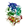
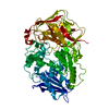
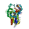
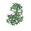
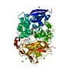
| Entry | Database: PDB / ID: 1l7q | ||||||
|---|---|---|---|---|---|---|---|
| Title | Ser117Ala Mutant of Bacterial Cocaine Esterase cocE | ||||||
 Components Components | cocaine esterase | ||||||
 Keywords Keywords | HYDROLASE / beta-alpha-beta / cocaine hydrolase / active site mutant / benzoate product complex | ||||||
| Function / homology |  Function and homology information Function and homology informationcocaine esterase / cocaine catabolic process / dipeptidyl-peptidase activity / carboxylic ester hydrolase activity / cytoplasm Similarity search - Function | ||||||
| Biological species |  Rhodococcus sp. MB1 (bacteria) Rhodococcus sp. MB1 (bacteria) | ||||||
| Method |  X-RAY DIFFRACTION / SYNCHROTRON / MOLECULAR REPLACEMENT / Resolution: 1.76 Å X-RAY DIFFRACTION / SYNCHROTRON / MOLECULAR REPLACEMENT / Resolution: 1.76 Å | ||||||
 Authors Authors | Turner, J.M. / Larsen, N.A. / Basran, A. / Barbas III, C.F. / Bruce, N.C. / Wilson, I.A. / Lerner, R.A. | ||||||
 Citation Citation | Journal: Biochemistry / Year: 2002 Title: Biochemical characterization and structural analysis of a highly proficient cocaine esterase Authors: Turner, J.M. / Larsen, N.A. / Basran, A. / Barbas III, C.F. / Bruce, N.C. / Wilson, I.A. / Lerner, R.A. | ||||||
| History |
|
- Structure visualization
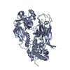
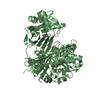
| Structure viewer | Molecule: MolmilJmol/JSmol |
|---|
- Downloads & links
-Download
| PDBx/mmCIF format | 1l7q.cif.gz | 134.4 KB | Display | PDBx/mmCIF format |
|---|---|---|---|---|
| PDB format | pdb1l7q.ent.gz | 103 KB | Display | PDB format |
| PDBx/mmJSON format | 1l7q.json.gz | Tree view | PDBx/mmJSON format | |
| Others |  Other downloads Other downloads |
-Validation report
| Arichive directory | https://data.pdbj.org/pub/pdb/validation_reports/l7/1l7qftp://data.pdbj.org/pub/pdb/validation_reports/l7/1l7q | HTTPS FTP |
|---|
-Related structure data
| Related structure data | 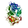 1l7rC 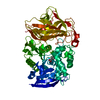 1ju4S S: Starting model for refinement C: citing same article ( |
|---|---|
| Similar structure data |


-Links
 PDBj
PDBj- Assembly
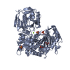
| Deposited unit | 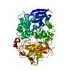
| ||||||||
|---|---|---|---|---|---|---|---|---|---|
| 1 |
| ||||||||
| Unit cell |
|
-Components
| #1: Protein | Mass: 62167.352 Da / Num. of mol.: 1 / Mutation: S117A Source method: isolated from a genetically manipulated source Source: (gene. exp.) Rhodococcus sp. MB1 (bacteria) / Production host: |
|---|---|
| #2: Chemical | ChemComp-BEZ /   Mass: 122.121 Da / Num. of mol.: 1 / Source method: obtained synthetically / Formula: C7H6O2 Mass: 122.121 Da / Num. of mol.: 1 / Source method: obtained synthetically / Formula: C7H6O2 |
| #3: Water | ChemComp-HOH /  Mass: 18.015 Da / Num. of mol.: 583 / Source method: isolated from a natural source / Formula: H2O Mass: 18.015 Da / Num. of mol.: 583 / Source method: isolated from a natural source / Formula: H2O |
-Experimental details
-Experiment
| Experiment | Method: X-RAY DIFFRACTION / Number of used crystals: 1 |
|---|
- Sample preparation
| Crystal | Density Matthews: 2.88 Å3/Da / Density % sol: 57.36 % | ||||||||||||||||||||||||||||||||||||||||||
|---|---|---|---|---|---|---|---|---|---|---|---|---|---|---|---|---|---|---|---|---|---|---|---|---|---|---|---|---|---|---|---|---|---|---|---|---|---|---|---|---|---|---|---|
| Crystal grow | Temperature: 296 K / Method: vapor diffusion, sitting drop / pH: 7.5 Details: 10 mM Tris, 25 mM NaCl, 1.4-1.6 M Ammonium Sulfate, pH 7.5, VAPOR DIFFUSION, SITTING DROP, temperature 296K | ||||||||||||||||||||||||||||||||||||||||||
| Crystal grow | *PLUS Method: microdialysis / Details: Larsen, N.A., (2002) Nature Struct. Biol., 9, 17. | ||||||||||||||||||||||||||||||||||||||||||
| Components of the solutions | *PLUS
|
-Data collection
| Diffraction | Mean temperature: 120 K |
|---|---|
| Diffraction source | Source: SYNCHROTRON / Site: SSRL  / Beamline: BL11-1 / Wavelength: 0.965 Å / Beamline: BL11-1 / Wavelength: 0.965 Å |
| Detector | Type: ADSC QUANTUM 4 / Detector: CCD / Date: May 31, 2001 |
| Radiation | Protocol: SINGLE WAVELENGTH / Monochromatic (M) / Laue (L): M / Scattering type: x-ray |
| Radiation wavelength | Wavelength: 0.965 Å / Relative weight: 1 |
| Reflection | Resolution: 1.76→30 Å / Num. all: 74054 / Num. obs: 74054 / % possible obs: 98.9 % / Observed criterion σ(F): 1.5 / Observed criterion σ(I): 2 / Rmerge(I) obs: 0.054 / Rsym value: 0.054 |
| Reflection shell | Resolution: 1.76→1.79 Å / Rmerge(I) obs: 0.494 / % possible all: 98.1 |
| Reflection | *PLUS Lowest resolution: 30 Å / Redundancy: 3.5 % |
| Reflection shell | *PLUS % possible obs: 98.1 % / Redundancy: 3.6 % / Mean I/σ(I) obs: 2.5 |
- Processing
| Software |
| ||||||||||||||||||||
|---|---|---|---|---|---|---|---|---|---|---|---|---|---|---|---|---|---|---|---|---|---|
| Refinement | Method to determine structure: MOLECULAR REPLACEMENT Starting model: 1JU4 Resolution: 1.76→30 Å / σ(F): 0 / Stereochemistry target values: Engh & Huber
| ||||||||||||||||||||
| Refine analyze |
| ||||||||||||||||||||
| Refinement step | Cycle: LAST / Resolution: 1.76→30 Å
| ||||||||||||||||||||
| Refine LS restraints |
| ||||||||||||||||||||
| Refinement | *PLUS Lowest resolution: 30 Å / % reflection Rfree: 5 % | ||||||||||||||||||||
| Solvent computation | *PLUS | ||||||||||||||||||||
| Displacement parameters | *PLUS | ||||||||||||||||||||
| Refine LS restraints | *PLUS
|
