 Movie
Movie Controller

[English] 日本語
 Yorodumi
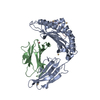
- PDB-4bc4: Crystal structure of human D-xylulokinase in complex with D-xylulose -
+ Open data

- Basic information
| Entry | Database: PDB / ID: 4bc4 | ||||||
|---|---|---|---|---|---|---|---|
| Title | Crystal structure of human D-xylulokinase in complex with D-xylulose | ||||||
 Components Components | XYLULOSE KINASE | ||||||
 Keywords Keywords | TRANSFERASE / GLUCURONATE XYLULOKINASE PATHWAY | ||||||
| Function / homology |  Function and homology information Function and homology informationxylulokinase / xylulose catabolic process / Formation of xylulose-5-phosphate / : / xylulose metabolic process / D-xylulokinase activity / D-xylose metabolic process / generation of precursor metabolites and energy / carbohydrate metabolic process / ATP binding / cytosol Similarity search - Function | ||||||
| Biological species |  HOMO SAPIENS (human) HOMO SAPIENS (human) | ||||||
| Method |  X-RAY DIFFRACTION / SYNCHROTRON / MOLECULAR REPLACEMENT / Resolution: 1.79 Å X-RAY DIFFRACTION / SYNCHROTRON / MOLECULAR REPLACEMENT / Resolution: 1.79 Å | ||||||
 Authors Authors | Bunker, R.D. / Loomes, K.M. / Baker, E.N. | ||||||
 Citation Citation | Journal: J.Biol.Chem. / Year: 2013 Title: Structure and Function of Human Xylulokinase, an Enzyme with Important Roles in Carbohydrate Metabolism Authors: Bunker, R.D. / Bulloch, E.M.M. / Dickson, J.M.J. / Loomes, K.M. / Baker, E.N. | ||||||
| History |
|
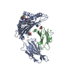
- Structure visualization
| Structure viewer | Molecule: MolmilJmol/JSmol |
|---|
- Downloads & links
-Download
| PDBx/mmCIF format | 4bc4.cif.gz | 1.1 MB | Display | PDBx/mmCIF format |
|---|---|---|---|---|
| PDB format | pdb4bc4.ent.gz | 950.6 KB | Display | PDB format |
| PDBx/mmJSON format | 4bc4.json.gz | Tree view | PDBx/mmJSON format | |
| Others |  Other downloads Other downloads |
-Validation report
| Arichive directory | https://data.pdbj.org/pub/pdb/validation_reports/bc/4bc4ftp://data.pdbj.org/pub/pdb/validation_reports/bc/4bc4 | HTTPS FTP |
|---|
-Related structure data
| Related structure data | 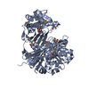 4bc2C  4bc3C  4bc5C  4b6t 4b6y C: citing same article ( |
|---|---|
| Similar structure data |


-Links
 PDBj
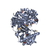
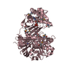
PDBj- Assembly
| Deposited unit | 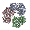
| ||||||||
|---|---|---|---|---|---|---|---|---|---|
| 1 | 
| ||||||||
| 2 | 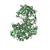
| ||||||||
| 3 | 
| ||||||||
| Unit cell |
|
-Components
| #1: Protein | Mass: 59175.680 Da / Num. of mol.: 3 Source method: isolated from a genetically manipulated source Source: (gene. exp.) HOMO SAPIENS (human) / Plasmid: PPROEX / Production host:  #2: Sugar | 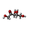  Type: D-saccharide / Mass: 150.130 Da / Num. of mol.: 3 Type: D-saccharide / Mass: 150.130 Da / Num. of mol.: 3Source method: isolated from a genetically manipulated source Formula: C5H10O5 #3: Chemical |   Mass: 62.068 Da / Num. of mol.: 3 / Source method: obtained synthetically / Formula: C2H6O2 Mass: 62.068 Da / Num. of mol.: 3 / Source method: obtained synthetically / Formula: C2H6O2#4: Water | ChemComp-HOH / |  Mass: 18.015 Da / Num. of mol.: 739 / Source method: isolated from a natural source / Formula: H2O Mass: 18.015 Da / Num. of mol.: 739 / Source method: isolated from a natural source / Formula: H2OHas protein modification | Y | |
|---|
-Experimental details
-Experiment
| Experiment | Method: X-RAY DIFFRACTION / Number of used crystals: 1 |
|---|
- Sample preparation
| Crystal | Density Matthews: 2.8 Å3/Da / Density % sol: 55 % / Description: NONE |
|---|---|
| Crystal grow | Details: 200 MM MES/KOH, PH 5.9, 15% PEG 6000 |
-Data collection
| Diffraction | Mean temperature: 100 K |
|---|---|
| Diffraction source | Source: SYNCHROTRON / Site: LNLS  / Beamline: W01B-MX2 / Wavelength: 0.8856 / Beamline: W01B-MX2 / Wavelength: 0.8856 |
| Detector | Type: MARRESEARCH / Detector: CCD / Date: Aug 13, 2009 / Details: MIRRORS |
| Radiation | Monochromator: SI(111) / Protocol: SINGLE WAVELENGTH / Monochromatic (M) / Laue (L): M / Scattering type: x-ray |
| Radiation wavelength | Wavelength: 0.8856 Å / Relative weight: 1 |
| Reflection | Resolution: 1.79→59.37 Å / Num. obs: 177198 / % possible obs: 99.8 % / Observed criterion σ(I): -3 / Redundancy: 11.3 % / Biso Wilson estimate: 30.02 Å2 / Rmerge(I) obs: 0.22 / Net I/σ(I): 9.7 |
| Reflection shell | Resolution: 1.79→1.82 Å / Redundancy: 10.1 % / Mean I/σ(I) obs: 0.5 / % possible all: 99.6 |
- Processing
| Software |
| ||||||||||||||||||||||||||||||||||||||||||||||||||||||||||||||||||||||||||||||||||||||||||||||||||||||||||||||||||
|---|---|---|---|---|---|---|---|---|---|---|---|---|---|---|---|---|---|---|---|---|---|---|---|---|---|---|---|---|---|---|---|---|---|---|---|---|---|---|---|---|---|---|---|---|---|---|---|---|---|---|---|---|---|---|---|---|---|---|---|---|---|---|---|---|---|---|---|---|---|---|---|---|---|---|---|---|---|---|---|---|---|---|---|---|---|---|---|---|---|---|---|---|---|---|---|---|---|---|---|---|---|---|---|---|---|---|---|---|---|---|---|---|---|---|---|
| Refinement | Method to determine structure: MOLECULAR REPLACEMENT Starting model: HUMAN D-XYLULOSE DETERMINED BY TWO-ENERGY SELENIUM MAD Resolution: 1.79→59.37 Å / Cor.coef. Fo:Fc: 0.9614 / Cor.coef. Fo:Fc free: 0.9556 / SU R Cruickshank DPI: 0.123 / Cross valid method: THROUGHOUT / σ(F): 0 / SU R Blow DPI: 0.093 / SU Rfree Blow DPI: 0.088 / SU Rfree Cruickshank DPI: 0.091
| ||||||||||||||||||||||||||||||||||||||||||||||||||||||||||||||||||||||||||||||||||||||||||||||||||||||||||||||||||
| Displacement parameters | Biso mean: 35.14 Å2
| ||||||||||||||||||||||||||||||||||||||||||||||||||||||||||||||||||||||||||||||||||||||||||||||||||||||||||||||||||
| Refine analyze | Luzzati coordinate error obs: 0.209 Å | ||||||||||||||||||||||||||||||||||||||||||||||||||||||||||||||||||||||||||||||||||||||||||||||||||||||||||||||||||
| Refinement step | Cycle: LAST / Resolution: 1.79→59.37 Å
| ||||||||||||||||||||||||||||||||||||||||||||||||||||||||||||||||||||||||||||||||||||||||||||||||||||||||||||||||||
| Refine LS restraints |
| ||||||||||||||||||||||||||||||||||||||||||||||||||||||||||||||||||||||||||||||||||||||||||||||||||||||||||||||||||
| LS refinement shell | Resolution: 1.79→1.84 Å / Total num. of bins used: 20
| ||||||||||||||||||||||||||||||||||||||||||||||||||||||||||||||||||||||||||||||||||||||||||||||||||||||||||||||||||
| Refinement TLS params. | Method: refined / Refine-ID: X-RAY DIFFRACTION
| ||||||||||||||||||||||||||||||||||||||||||||||||||||||||||||||||||||||||||||||||||||||||||||||||||||||||||||||||||
| Refinement TLS group |
|
