Journal: AM.CRYST.ASSOC.,ABSTR.PAPERS (ANNUAL MEETING) / Year: 2002 Title: Structure determination of P. aeruginosa lectin-1 using single wavelength anomalous scattering data from native crystals (P028) Authors: Liu, Z.J. / Tempel, W. / Lin, D. / Karaveg, K. / Doyle, R.J. / Rose, J.P. / Wang, B.C.
Resolution: 1.5→1.55 Å / Redundancy: 4.2 % / Rmerge(I) obs: 0.14 / Mean I/σ(I) obs: 10 / Num. unique all: 3029 / Rsym value: 0.14 / % possible all: 96.3
-
Processing
Software
Name
Version
Classification
REFMAC
5.1.24
refinement
SCALEPACK
datascaling
SHELX
modelbuilding
ISAS
modelbuilding
RESOLVE
modelbuilding
CNS
1.1
refinement
SHELX
phasing
ISAS
phasing
RESOLVE
phasing
MAR345
datacollection
Refinement
Method to determine structure: SAS / Resolution: 1.5→14.6 Å / Cor.coef. Fo:Fc: 0.947 / Cor.coef. Fo:Fc free: 0.947 / SU B: 0.793 / SU ML: 0.031 / Cross valid method: THROUGHOUT / ESU R: 0.059 / ESU R Free: 0.057 / Stereochemistry target values: MAXIMUM LIKELIHOOD / Details: HYDROGENS HAVE BEEN ADDED IN THE RIDING POSITIONS
Rfactor
Num. reflection
% reflection
Selection details
Rfree
0.2002
1535
5 %
RANDOM
Rwork
0.19225
-
-
-
obs
0.19262
29435
97.31 %
-
Solvent computation
Ion probe radii: 0.8 Å / Shrinkage radii: 0.8 Å / VDW probe radii: 1.4 Å / Solvent model: BABINET MODEL WITH MASK
Displacement parameters
Biso mean: 13.435 Å2
Baniso -1
Baniso -2
Baniso -3
1-
1.14 Å2
0 Å2
0 Å2
2-
-
-0.19 Å2
0 Å2
3-
-
-
-0.94 Å2
Refine analyze
Free
Obs
Luzzati coordinate error
0.18 Å
0.17 Å
Luzzati d res low
-
5 Å
Luzzati sigma a
0.06 Å
-0.04 Å
Refinement step
Cycle: LAST / Resolution: 1.5→14.6 Å
Protein
Nucleic acid
Ligand
Solvent
Total
Num. atoms
897
0
1
107
1005
Refine LS restraints
Refine-ID
Type
Dev ideal
Dev ideal target
Number
X-RAY DIFFRACTION
r_bond_refined_d
0.011
0.021
920
X-RAY DIFFRACTION
r_bond_other_d
0.002
0.02
788
X-RAY DIFFRACTION
r_angle_refined_deg
1.301
1.916
1257
X-RAY DIFFRACTION
r_angle_other_deg
0.722
3
1840
X-RAY DIFFRACTION
r_dihedral_angle_1_deg
5.971
5
120
X-RAY DIFFRACTION
r_dihedral_angle_2_deg
X-RAY DIFFRACTION
r_chiral_restr
0.082
0.2
135
X-RAY DIFFRACTION
r_gen_planes_refined
0.006
0.02
1066
X-RAY DIFFRACTION
r_gen_planes_other
0.004
0.02
176
X-RAY DIFFRACTION
r_nbd_refined
0.2
0.2
145
X-RAY DIFFRACTION
r_nbd_other
0.252
0.2
874
X-RAY DIFFRACTION
r_nbtor_other
0.083
0.2
514
X-RAY DIFFRACTION
r_xyhbond_nbd_refined
0.092
0.2
58
X-RAY DIFFRACTION
r_metal_ion_refined
0.062
0.2
3
X-RAY DIFFRACTION
r_xyhbond_nbd_other
X-RAY DIFFRACTION
r_symmetry_vdw_refined
0.152
0.2
5
X-RAY DIFFRACTION
r_symmetry_vdw_other
0.327
0.2
33
X-RAY DIFFRACTION
r_symmetry_hbond_refined
0.129
0.2
8
X-RAY DIFFRACTION
r_symmetry_hbond_other
X-RAY DIFFRACTION
r_mcbond_it
0.737
1.5
594
X-RAY DIFFRACTION
r_mcangle_it
1.316
2
950
X-RAY DIFFRACTION
r_scbond_it
2.169
3
326
X-RAY DIFFRACTION
r_scangle_it
3.352
4.5
307
X-RAY DIFFRACTION
r_rigid_bond_restr
X-RAY DIFFRACTION
r_sphericity_free
X-RAY DIFFRACTION
r_sphericity_bonded
LS refinement shell
Resolution: 1.5→1.539 Å / Total num. of bins used: 20 /
Rfactor
Num. reflection
Rfree
0.207
124
Rwork
0.184
2025
Xplor file
Refine-ID
Serial no
Param file
Topol file
X-RAY DIFFRACTION
1
PROTEIN_REP.PARAM
PROTEIN.TOP
X-RAY DIFFRACTION
2
WATER_REP.PARAM
WATER.TOP
X-RAY DIFFRACTION
3
ION.PARAM
ION.TOP
+
About Yorodumi
-
News
-
Feb 9, 2022. New format data for meta-information of EMDB entries
New format data for meta-information of EMDB entries
Version 3 of the EMDB header file is now the official format.
The previous official version 1.9 will be removed from the archive.
In the structure databanks used in Yorodumi, some data are registered as the other names, "COVID-19 virus" and "2019-nCoV". Here are the details of the virus and the list of structure data.
Jan 31, 2019. EMDB accession codes are about to change! (news from PDBe EMDB page)
EMDB accession codes are about to change! (news from PDBe EMDB page)
The allocation of 4 digits for EMDB accession codes will soon come to an end. Whilst these codes will remain in use, new EMDB accession codes will include an additional digit and will expand incrementally as the available range of codes is exhausted. The current 4-digit format prefixed with “EMD-” (i.e. EMD-XXXX) will advance to a 5-digit format (i.e. EMD-XXXXX), and so on. It is currently estimated that the 4-digit codes will be depleted around Spring 2019, at which point the 5-digit format will come into force.
The EM Navigator/Yorodumi systems omit the EMD- prefix.
Related info.:Q: What is EMD? / ID/Accession-code notation in Yorodumi/EM Navigator
Yorodumi is a browser for structure data from EMDB, PDB, SASBDB, etc.
This page is also the successor to EM Navigator detail page, and also detail information page/front-end page for Omokage search.
The word "yorodu" (or yorozu) is an old Japanese word meaning "ten thousand". "mi" (miru) is to see.
Related info.:EMDB / PDB / SASBDB / Comparison of 3 databanks / Yorodumi Search / Aug 31, 2016. New EM Navigator & Yorodumi / Yorodumi Papers / Jmol/JSmol / Function and homology information / Changes in new EM Navigator and Yorodumi
 Movie
Movie Controller

 Yorodumi
 Open data
 Basic information
 Components
 Keywords
 Function and homology information
 Pseudomonas aeruginosa (bacteria)
 X-RAY DIFFRACTION /
  Authors
 Citation
 Structure visualization
 Downloads & links
 Other downloads


 PDBj
PDBj Assembly
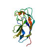
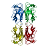

 Mass: 40.078 Da / Num. of mol.: 1 / Source method: obtained synthetically / Formula: Ca
 Mass: 18.015 Da / Num. of mol.: 107 / Source method: isolated from a natural source / Formula: H2O
 Sample preparation
 / Beamline: 17-ID / Wavelength: 1 Å
 Processing