 Movie
Movie Controller

[English] 日本語
 Yorodumi
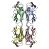
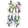
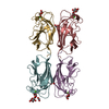
- PDB-1uoj: CRYSTAL STRUCTURE OF PSEUDOMONAS AERUGINOSA LECTIN 1 IN THE CALCI... -
+ Open data

- Basic information
| Entry | Database: PDB / ID: 1uoj | ||||||
|---|---|---|---|---|---|---|---|
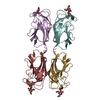
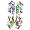
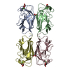
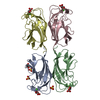
| Title | CRYSTAL STRUCTURE OF PSEUDOMONAS AERUGINOSA LECTIN 1 IN THE CALCIUM-FREE STATE | ||||||
 Components Components | PA-I GALACTOPHILIC LECTIN | ||||||
 Keywords Keywords | SUGAR BINDING PROTEIN / GALACTOSE BINDING | ||||||
| Function / homology |  Function and homology information Function and homology informationheterophilic cell-cell adhesion / carbohydrate binding / periplasmic space / cell surface / cytoplasm Similarity search - Function | ||||||
| Biological species |   PSEUDOMONAS AERUGINOSA (bacteria) PSEUDOMONAS AERUGINOSA (bacteria) | ||||||
| Method |  X-RAY DIFFRACTION / SYNCHROTRON / MOLECULAR REPLACEMENT / Resolution: 2.4 Å X-RAY DIFFRACTION / SYNCHROTRON / MOLECULAR REPLACEMENT / Resolution: 2.4 Å | ||||||
 Authors Authors | Cioci, G. / Mitchell, E. / Gautier, C. / Wimmerova, M. / Perez, S. / Gilboa-Garber, N. / Imberty, A. | ||||||
 Citation Citation | Journal: FEBS Lett. / Year: 2003 Title: Structural Basis of Calcium and Galactose Recognition by the Lectin Pa-Il of Pseudomonas Aeruginosa Authors: Cioci, G. / Mitchell, E. / Gautier, C. / Wimmerova, M. / Sudakevitz, D. / Perez, S. / Gilboa-Garber, N. / Imberty, A. | ||||||
| History |
| ||||||
| Remark 700 | SHEET THE SHEET STRUCTURE OF THIS MOLECULE IS BIFURCATED. IN ORDER TO REPRESENT THIS FEATURE IN ... SHEET THE SHEET STRUCTURE OF THIS MOLECULE IS BIFURCATED. IN ORDER TO REPRESENT THIS FEATURE IN THE SHEET RECORDS BELOW, TWO SHEETS ARE DEFINED. |
- Structure visualization
| Structure viewer | Molecule: MolmilJmol/JSmol |
|---|
- Downloads & links
-Download
| PDBx/mmCIF format | 1uoj.cif.gz | 101.8 KB | Display | PDBx/mmCIF format |
|---|---|---|---|---|
| PDB format | pdb1uoj.ent.gz | 79.1 KB | Display | PDB format |
| PDBx/mmJSON format | 1uoj.json.gz | Tree view | PDBx/mmJSON format | |
| Others |  Other downloads Other downloads |
-Validation report
| Arichive directory | https://data.pdbj.org/pub/pdb/validation_reports/uo/1uojftp://data.pdbj.org/pub/pdb/validation_reports/uo/1uoj | HTTPS FTP |
|---|
-Related structure data
| Related structure data |  1okoC 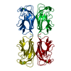 1l7lS S: Starting model for refinement C: citing same article ( |
|---|---|
| Similar structure data |


-Links
 PDBj
PDBj- Assembly
| Deposited unit | 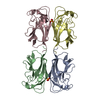
| ||||||||
|---|---|---|---|---|---|---|---|---|---|
| 1 |
| ||||||||
| Unit cell |
|
-Components
| #1: Protein | Mass: 12770.137 Da / Num. of mol.: 4 / Source method: isolated from a natural source / Source: (natural) PSEUDOMONAS AERUGINOSA (bacteria) / References: UniProt: Q05097#2: Chemical |   Mass: 96.063 Da / Num. of mol.: 2 / Source method: obtained synthetically / Formula: SO4 Mass: 96.063 Da / Num. of mol.: 2 / Source method: obtained synthetically / Formula: SO4#3: Water | ChemComp-HOH / |  Mass: 18.015 Da / Num. of mol.: 137 / Source method: isolated from a natural source / Formula: H2O Mass: 18.015 Da / Num. of mol.: 137 / Source method: isolated from a natural source / Formula: H2OCompound details | D-GALACTOSE SPECIFIC LECTIN THAT BINDS IN DECREASING ORDER OF AFFINITY: MELIBIOSE, METHYL-ALPHA-D- ...D-GALACTOSE SPECIFIC LECTIN THAT BINDS IN DECREASING | |
|---|
-Experimental details
-Experiment
| Experiment | Method: X-RAY DIFFRACTION / Number of used crystals: 1 |
|---|
- Sample preparation
| Crystal | Density Matthews: 2 Å3/Da / Density % sol: 37 % | ||||||||||||||||||||||||
|---|---|---|---|---|---|---|---|---|---|---|---|---|---|---|---|---|---|---|---|---|---|---|---|---|---|
| Crystal grow | Method: vapor diffusion, hanging drop / pH: 5 Details: HANGING DROP: PROTEIN SOLUTION: PA1L 10 MG/ML + DGAL 0.5 MG/ML. RESERVOIR SOLUTION (NH4)2SO4, 1.5 M 20% ISOPROPANOL PH 4.5 2 UL + 2 UL | ||||||||||||||||||||||||
| Crystal grow | *PLUS pH: 4.5 / Method: vapor diffusion, hanging drop | ||||||||||||||||||||||||
| Components of the solutions | *PLUS
|
-Data collection
| Diffraction | Mean temperature: 100 K |
|---|---|
| Diffraction source | Source: SYNCHROTRON / Site: ESRF  / Beamline: ID14-4 / Wavelength: 0.932 / Beamline: ID14-4 / Wavelength: 0.932 |
| Detector | Type: ADSC CCD / Detector: CCD / Date: Mar 15, 2003 / Details: MIRROR |
| Radiation | Monochromator: SILICON / Protocol: SINGLE WAVELENGTH / Monochromatic (M) / Laue (L): M / Scattering type: x-ray |
| Radiation wavelength | Wavelength: 0.932 Å / Relative weight: 1 |
| Reflection | Resolution: 2.4→20.37 Å / Num. obs: 17328 / % possible obs: 99.8 % / Redundancy: 6.54 % / Rmerge(I) obs: 0.089 / Net I/σ(I): 7.6715 |
| Reflection shell | Resolution: 2.4→2.48 Å / Redundancy: 6.19 % / Rmerge(I) obs: 0.36 / Mean I/σ(I) obs: 1.93 / % possible all: 99.8 |
| Reflection | *PLUS Highest resolution: 2.4 Å / Num. obs: 17451 / Redundancy: 6.5 % / Num. measured all: 114114 / Rmerge(I) obs: 0.089 |
| Reflection shell | *PLUS % possible obs: 99.8 % / Redundancy: 6.2 % / Rmerge(I) obs: 0.36 / Mean I/σ(I) obs: 1.9 |
- Processing
| Software |
| ||||||||||||||||||||||||||||||||||||||||||||||||||||||||||||||||||||||||||||||||||||||||||||||||||||||||||||||||||||||||||||||||||||||||||||||||||||||||||||||||||||||||||||||||||||||
|---|---|---|---|---|---|---|---|---|---|---|---|---|---|---|---|---|---|---|---|---|---|---|---|---|---|---|---|---|---|---|---|---|---|---|---|---|---|---|---|---|---|---|---|---|---|---|---|---|---|---|---|---|---|---|---|---|---|---|---|---|---|---|---|---|---|---|---|---|---|---|---|---|---|---|---|---|---|---|---|---|---|---|---|---|---|---|---|---|---|---|---|---|---|---|---|---|---|---|---|---|---|---|---|---|---|---|---|---|---|---|---|---|---|---|---|---|---|---|---|---|---|---|---|---|---|---|---|---|---|---|---|---|---|---|---|---|---|---|---|---|---|---|---|---|---|---|---|---|---|---|---|---|---|---|---|---|---|---|---|---|---|---|---|---|---|---|---|---|---|---|---|---|---|---|---|---|---|---|---|---|---|---|---|
| Refinement | Method to determine structure: MOLECULAR REPLACEMENT Starting model: PDB ENTRY 1L7L Resolution: 2.4→20.37 Å / Cor.coef. Fo:Fc: 0.943 / Cor.coef. Fo:Fc free: 0.904 / SU B: 8.76 / SU ML: 0.205 / Cross valid method: THROUGHOUT / ESU R: 0.691 / ESU R Free: 0.306 / Stereochemistry target values: MAXIMUM LIKELIHOOD / Details: HYDROGENS HAVE BEEN ADDED IN THE RIDING POSITIONS
| ||||||||||||||||||||||||||||||||||||||||||||||||||||||||||||||||||||||||||||||||||||||||||||||||||||||||||||||||||||||||||||||||||||||||||||||||||||||||||||||||||||||||||||||||||||||
| Solvent computation | Ion probe radii: 0.8 Å / Shrinkage radii: 0.8 Å / VDW probe radii: 1.4 Å / Solvent model: BABINET MODEL PLUS MASK | ||||||||||||||||||||||||||||||||||||||||||||||||||||||||||||||||||||||||||||||||||||||||||||||||||||||||||||||||||||||||||||||||||||||||||||||||||||||||||||||||||||||||||||||||||||||
| Displacement parameters | Biso mean: 27.72 Å2
| ||||||||||||||||||||||||||||||||||||||||||||||||||||||||||||||||||||||||||||||||||||||||||||||||||||||||||||||||||||||||||||||||||||||||||||||||||||||||||||||||||||||||||||||||||||||
| Refinement step | Cycle: LAST / Resolution: 2.4→20.37 Å
| ||||||||||||||||||||||||||||||||||||||||||||||||||||||||||||||||||||||||||||||||||||||||||||||||||||||||||||||||||||||||||||||||||||||||||||||||||||||||||||||||||||||||||||||||||||||
| Refine LS restraints |
|