 Movie
Movie Controller

[English] 日本語
 Yorodumi
- PDB-6yo3: LecA from Pseudomonas aeruginosa in complex with a catechol CAS n... -
+ Open data

- Basic information
| Entry | Database: PDB / ID: 6yo3 | |||||||||||||||||||||
|---|---|---|---|---|---|---|---|---|---|---|---|---|---|---|---|---|---|---|---|---|---|---|
| Title | LecA from Pseudomonas aeruginosa in complex with a catechol CAS no. 67984-81-0 | |||||||||||||||||||||
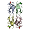
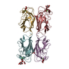
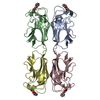
 Components Components | PA-I galactophilic lectin | |||||||||||||||||||||
 Keywords Keywords | SUGAR BINDING PROTEIN / Non-carbohydrate glycomimetics / PAINS / lectin / catechols | |||||||||||||||||||||
| Function / homology |  Function and homology information Function and homology informationheterophilic cell-cell adhesion / carbohydrate binding / periplasmic space / cell surface / cytoplasm Similarity search - Function | |||||||||||||||||||||
| Biological species |   Pseudomonas aeruginosa (bacteria) Pseudomonas aeruginosa (bacteria) | |||||||||||||||||||||
| Method |  X-RAY DIFFRACTION / SYNCHROTRON / MOLECULAR REPLACEMENT / Resolution: 1.84 Å X-RAY DIFFRACTION / SYNCHROTRON / MOLECULAR REPLACEMENT / Resolution: 1.84 Å | |||||||||||||||||||||
 Authors Authors | Kuhaudomlarp, S. / Imberty, A. / Titz, A. | |||||||||||||||||||||
| Funding support |  France, France,  Germany, 6items Germany, 6items
| |||||||||||||||||||||
 Citation Citation | Journal: Angew.Chem.Int.Ed.Engl. / Year: 2021 Title: Non-Carbohydrate Glycomimetics as Inhibitors of Calcium(II)-Binding Lectins. Authors: Kuhaudomlarp, S. / Siebs, E. / Shanina, E. / Topin, J. / Joachim, I. / da Silva Figueiredo Celestino Gomes, P. / Varrot, A. / Rognan, D. / Rademacher, C. / Imberty, A. / Titz, A. | |||||||||||||||||||||
| History |
|
- Structure visualization
| Structure viewer | Molecule: MolmilJmol/JSmol |
|---|
- Downloads & links
-Download
| PDBx/mmCIF format | 6yo3.cif.gz | 114.9 KB | Display | PDBx/mmCIF format |
|---|---|---|---|---|
| PDB format | pdb6yo3.ent.gz | 87.1 KB | Display | PDB format |
| PDBx/mmJSON format | 6yo3.json.gz | Tree view | PDBx/mmJSON format | |
| Others |  Other downloads Other downloads |
-Validation report
| Arichive directory | https://data.pdbj.org/pub/pdb/validation_reports/yo/6yo3ftp://data.pdbj.org/pub/pdb/validation_reports/yo/6yo3 | HTTPS FTP |
|---|
-Related structure data
| Related structure data | 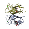 6yohC  1okoS S: Starting model for refinement C: citing same article ( |
|---|---|
| Similar structure data |


-Links
 PDBj
PDBj- Assembly
| Deposited unit | 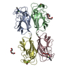
| ||||||||
|---|---|---|---|---|---|---|---|---|---|
| 1 |
| ||||||||
| Unit cell |
|
-Components
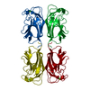
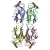
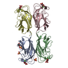
-Protein , 1 types, 4 molecules ABCD
| #1: Protein | Mass: 12770.137 Da / Num. of mol.: 4 Source method: isolated from a genetically manipulated source Details: LecA from Pseudomonas aeruginosa was produced as a recombinant protein in E. coli Source: (gene. exp.) Pseudomonas aeruginosa (strain ATCC 15692 / DSM 22644 / CIP 104116 / JCM 14847 / LMG 12228 / 1C / PRS 101 / PAO1) (bacteria)Strain: ATCC 15692 / DSM 22644 / CIP 104116 / JCM 14847 / LMG 12228 / 1C / PRS 101 / PAO1 Gene: lecA, pa1L, PA2570 / Production host: |
|---|
-Non-polymers , 7 types, 364 molecules 
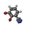


| #2: Chemical | ChemComp-CA /  Mass: 40.078 Da / Num. of mol.: 4 / Source method: obtained synthetically / Formula: Ca Mass: 40.078 Da / Num. of mol.: 4 / Source method: obtained synthetically / Formula: Ca#3: Chemical |  Mass: 135.120 Da / Num. of mol.: 2 / Source method: obtained synthetically / Formula: C7H5NO2 / Feature type: SUBJECT OF INVESTIGATION Mass: 135.120 Da / Num. of mol.: 2 / Source method: obtained synthetically / Formula: C7H5NO2 / Feature type: SUBJECT OF INVESTIGATION#4: Chemical | ChemComp-P6G / |  Mass: 282.331 Da / Num. of mol.: 1 / Source method: obtained synthetically / Formula: C12H26O7 / Comment: precipitant*YM Mass: 282.331 Da / Num. of mol.: 1 / Source method: obtained synthetically / Formula: C12H26O7 / Comment: precipitant*YM#5: Chemical |  Mass: 62.068 Da / Num. of mol.: 2 / Source method: obtained synthetically / Formula: C2H6O2 Mass: 62.068 Da / Num. of mol.: 2 / Source method: obtained synthetically / Formula: C2H6O2#6: Chemical | ChemComp-PG4 / |  Mass: 194.226 Da / Num. of mol.: 1 / Source method: obtained synthetically / Formula: C8H18O5 / Comment: precipitant*YM Mass: 194.226 Da / Num. of mol.: 1 / Source method: obtained synthetically / Formula: C8H18O5 / Comment: precipitant*YM#7: Chemical | ChemComp-PGE / |  Mass: 150.173 Da / Num. of mol.: 1 / Source method: obtained synthetically / Formula: C6H14O4 Mass: 150.173 Da / Num. of mol.: 1 / Source method: obtained synthetically / Formula: C6H14O4#8: Water | ChemComp-HOH / | Mass: 18.015 Da / Num. of mol.: 353 / Source method: isolated from a natural source / Formula: H2O |
|---|
-Details
| Has ligand of interest | Y |
|---|
-Experimental details
-Experiment
| Experiment | Method: X-RAY DIFFRACTION / Number of used crystals: 1 |
|---|
- Sample preparation
| Crystal | Density Matthews: 2.71 Å3/Da / Density % sol: 54.61 % |
|---|---|
| Crystal grow | Temperature: 292.15 K / Method: vapor diffusion, hanging drop / pH: 4.5 Details: 20% PEG6000, 100 mM sodium acetate pH 4.5, 1 M LiCl, 1% DMSO mixed in 1:1 ratio with 10mg/ml of LecA in water containing 100 uM CaCl2. The mixture was deposited onto dried catechol compound ...Details: 20% PEG6000, 100 mM sodium acetate pH 4.5, 1 M LiCl, 1% DMSO mixed in 1:1 ratio with 10mg/ml of LecA in water containing 100 uM CaCl2. The mixture was deposited onto dried catechol compound for co-crystallisation |
-Data collection
| Diffraction | Mean temperature: 100 K / Serial crystal experiment: N | |||||||||||||||||||||||||||
|---|---|---|---|---|---|---|---|---|---|---|---|---|---|---|---|---|---|---|---|---|---|---|---|---|---|---|---|---|
| Diffraction source | Source: SYNCHROTRON / Site: SOLEIL / Beamline: PROXIMA 1 / Wavelength: 0.9786 Å | |||||||||||||||||||||||||||
| Detector | Type: DECTRIS PILATUS 6M / Detector: PIXEL / Date: Jul 25, 2019 | |||||||||||||||||||||||||||
| Radiation | Protocol: SINGLE WAVELENGTH / Monochromatic (M) / Laue (L): M / Scattering type: x-ray | |||||||||||||||||||||||||||
| Radiation wavelength | Wavelength: 0.9786 Å / Relative weight: 1 | |||||||||||||||||||||||||||
| Reflection | Resolution: 1.84→45.5 Å / Num. obs: 47830 / % possible obs: 99.6 % / Redundancy: 5.6 % / CC1/2: 0.997 / Rmerge(I) obs: 0.09 / Rpim(I) all: 0.041 / Rrim(I) all: 0.099 / Net I/σ(I): 11.1 | |||||||||||||||||||||||||||
| Reflection shell | Diffraction-ID: 1 / Redundancy: 5.4 %
|
- Processing
| Software |
| ||||||||||||||||||||||||||||||||||||||||||||||||||||||||||||
|---|---|---|---|---|---|---|---|---|---|---|---|---|---|---|---|---|---|---|---|---|---|---|---|---|---|---|---|---|---|---|---|---|---|---|---|---|---|---|---|---|---|---|---|---|---|---|---|---|---|---|---|---|---|---|---|---|---|---|---|---|---|
| Refinement | Method to determine structure: MOLECULAR REPLACEMENT Starting model: 1OKO Resolution: 1.84→45.46 Å / Cor.coef. Fo:Fc: 0.961 / Cor.coef. Fo:Fc free: 0.94 / SU B: 2.711 / SU ML: 0.08 / SU R Cruickshank DPI: 0.1208 / Cross valid method: THROUGHOUT / σ(F): 0 / ESU R: 0.121 / ESU R Free: 0.117 Details: HYDROGENS HAVE BEEN ADDED IN THE RIDING POSITIONS U VALUES : REFINED INDIVIDUALLY
| ||||||||||||||||||||||||||||||||||||||||||||||||||||||||||||
| Solvent computation | Ion probe radii: 0.8 Å / Shrinkage radii: 0.8 Å / VDW probe radii: 1.2 Å | ||||||||||||||||||||||||||||||||||||||||||||||||||||||||||||
| Displacement parameters | Biso max: 65.6 Å2 / Biso mean: 21.456 Å2 / Biso min: 8.5 Å2
| ||||||||||||||||||||||||||||||||||||||||||||||||||||||||||||
| Refinement step | Cycle: final / Resolution: 1.84→45.46 Å
| ||||||||||||||||||||||||||||||||||||||||||||||||||||||||||||
| Refine LS restraints |
| ||||||||||||||||||||||||||||||||||||||||||||||||||||||||||||
| LS refinement shell | Resolution: 1.84→1.885 Å / Rfactor Rfree error: 0
|
