 Movie
Movie Controller


+ Open data

- Basic information
| Entry | Database: PDB / ID: 1l6w | ||||||
|---|---|---|---|---|---|---|---|
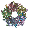
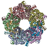
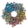
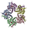
| Title | Fructose-6-phosphate aldolase | ||||||
 Components Components | Fructose-6-phosphate aldolase 1 | ||||||
 Keywords Keywords | LYASE / alpha-beta barrel / domain swapping | ||||||
| Function / homology |  Function and homology information Function and homology informationketone catabolic process / fructose 6-phosphate aldolase activity / Lyases; Carbon-carbon lyases; Aldehyde-lyases / fructose metabolic process / identical protein binding / cytoplasm Similarity search - Function | ||||||
| Biological species |  | ||||||
| Method |  X-RAY DIFFRACTION / SYNCHROTRON / SIR / Resolution: 1.93 Å X-RAY DIFFRACTION / SYNCHROTRON / SIR / Resolution: 1.93 Å | ||||||
 Authors Authors | Thorell, S. / Schuermann, M. / Sprenger, G.A. / Schneider, G. | ||||||
 Citation Citation | Journal: J.Mol.Biol. / Year: 2002 Title: Crystal structure of decameric fructose-6-phosphate aldolase from Escherichia coli reveals inter-subunit helix swapping as a structural basis for assembly differences in the transaldolase family. Authors: Thorell, S. / Schurmann, M. / Sprenger, G.A. / Schneider, G. #1: Journal: J.Biol.Chem. / Year: 2000Title: Fructose-6-phosphate Aldolase Is a Novel Class I Aldolase from Escherichia coli and Is Related to a Novel Group of Bacterial Transaldolases Authors: Schuermann, M. / Sprenger, G.A. | ||||||
| History |
|
- Structure visualization
| Structure viewer | Molecule: MolmilJmol/JSmol |
|---|
- Downloads & links
-Download
| PDBx/mmCIF format | 1l6w.cif.gz | 427 KB | Display | PDBx/mmCIF format |
|---|---|---|---|---|
| PDB format | pdb1l6w.ent.gz | 349.8 KB | Display | PDB format |
| PDBx/mmJSON format | 1l6w.json.gz | Tree view | PDBx/mmJSON format | |
| Others |  Other downloads Other downloads |
-Validation report
| Arichive directory | https://data.pdbj.org/pub/pdb/validation_reports/l6/1l6wftp://data.pdbj.org/pub/pdb/validation_reports/l6/1l6w | HTTPS FTP |
|---|
-Related structure data
| Related structure data | |
|---|---|
| Similar structure data |


-Links
 PDBj
PDBj- Assembly
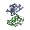
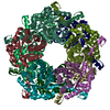
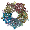
| Deposited unit | 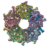
| ||||||||
|---|---|---|---|---|---|---|---|---|---|
| 1 |
| ||||||||
| Unit cell |
|
-Components
| #1: Protein | Mass: 23016.756 Da / Num. of mol.: 10 Source method: isolated from a genetically manipulated source Source: (gene. exp.) References: UniProt: P78055, Lyases; Carbon-carbon lyases; Aldehyde-lyases #2: Chemical | ChemComp-GOL /   Mass: 92.094 Da / Num. of mol.: 10 / Source method: obtained synthetically / Formula: C3H8O3 Mass: 92.094 Da / Num. of mol.: 10 / Source method: obtained synthetically / Formula: C3H8O3#3: Water | ChemComp-HOH / |  Mass: 18.015 Da / Num. of mol.: 1479 / Source method: isolated from a natural source / Formula: H2O Mass: 18.015 Da / Num. of mol.: 1479 / Source method: isolated from a natural source / Formula: H2OHas protein modification | Y | |
|---|
-Experimental details
-Experiment
| Experiment | Method: X-RAY DIFFRACTION / Number of used crystals: 1 |
|---|
- Sample preparation
| Crystal | Density Matthews: 2.92 Å3/Da / Density % sol: 57.91 % | ||||||||||||||||||||||||||||||||||||||||||
|---|---|---|---|---|---|---|---|---|---|---|---|---|---|---|---|---|---|---|---|---|---|---|---|---|---|---|---|---|---|---|---|---|---|---|---|---|---|---|---|---|---|---|---|
| Crystal grow | Temperature: 298 K / Method: vapor diffusion, hanging drop / pH: 4.6 Details: PEG4000, sodium acetate, glycerol, pH 4.6, VAPOR DIFFUSION, HANGING DROP, temperature 298.0K | ||||||||||||||||||||||||||||||||||||||||||
| Crystal grow | *PLUS Temperature: 20 ℃ / pH: 8 / Method: vapor diffusion | ||||||||||||||||||||||||||||||||||||||||||
| Components of the solutions | *PLUS
|
-Data collection
| Diffraction | Mean temperature: 107 K |
|---|---|
| Diffraction source | Source: SYNCHROTRON / Site: MAX II  / Beamline: I711 / Beamline: I711 |
| Detector | Type: MARRESEARCH / Detector: IMAGE PLATE / Date: Feb 5, 2000 |
| Radiation | Protocol: SINGLE WAVELENGTH / Monochromatic (M) / Laue (L): M / Scattering type: x-ray |
| Radiation wavelength | Relative weight: 1 |
| Reflection | Resolution: 1.93→19.94 Å / Num. all: 200850 / Num. obs: 200177 / % possible obs: 99.1 % / Observed criterion σ(F): 0 / Observed criterion σ(I): 0 / Redundancy: 7 % / Biso Wilson estimate: 11.3 Å2 / Limit h max: 59 / Limit h min: 0 / Limit k max: 65 / Limit k min: 0 / Limit l max: 95 / Limit l min: 0 / Observed criterion F max: 3950506.21 / Observed criterion F min: 11 / Rmerge(I) obs: 0.038 / Net I/σ(I): 46 |
| Reflection shell | Resolution: 1.93→1.96 Å / Rmerge(I) obs: 0.125 / Mean I/σ(I) obs: 18 / % possible all: 100 |
| Reflection | *PLUS Lowest resolution: 20 Å / Num. obs: 200850 / Rmerge(I) obs: 0.038 |
| Reflection shell | *PLUS % possible obs: 100 % / Rmerge(I) obs: 0.125 / Mean I/σ(I) obs: 18 |
- Processing
| Software |
| |||||||||||||||||||||||||||||||||||||||||||||||||||||||||||||||||||||||||||||||||
|---|---|---|---|---|---|---|---|---|---|---|---|---|---|---|---|---|---|---|---|---|---|---|---|---|---|---|---|---|---|---|---|---|---|---|---|---|---|---|---|---|---|---|---|---|---|---|---|---|---|---|---|---|---|---|---|---|---|---|---|---|---|---|---|---|---|---|---|---|---|---|---|---|---|---|---|---|---|---|---|---|---|---|
| Refinement | Method to determine structure: SIR / Resolution: 1.93→19.94 Å / Rfactor Rfree error: 0.002 / Occupancy max: 1 / Occupancy min: 0 / Isotropic thermal model: restrained / Cross valid method: THROUGHOUT / σ(F): 0 / Stereochemistry target values: Engh & Huber
| |||||||||||||||||||||||||||||||||||||||||||||||||||||||||||||||||||||||||||||||||
| Solvent computation | Solvent model: CNS bulk solvent model used / Bsol: 60.0517 Å2 / ksol: 0.412232 e/Å3 | |||||||||||||||||||||||||||||||||||||||||||||||||||||||||||||||||||||||||||||||||
| Displacement parameters | Biso max: 61.2 Å2 / Biso mean: 18.93 Å2 / Biso min: 6.63 Å2
| |||||||||||||||||||||||||||||||||||||||||||||||||||||||||||||||||||||||||||||||||
| Refine Biso |
| |||||||||||||||||||||||||||||||||||||||||||||||||||||||||||||||||||||||||||||||||
| Refine analyze |
| |||||||||||||||||||||||||||||||||||||||||||||||||||||||||||||||||||||||||||||||||
| Refinement step | Cycle: LAST / Resolution: 1.93→19.94 Å
| |||||||||||||||||||||||||||||||||||||||||||||||||||||||||||||||||||||||||||||||||
| Refine LS restraints |
| |||||||||||||||||||||||||||||||||||||||||||||||||||||||||||||||||||||||||||||||||
| LS refinement shell | Refine-ID: X-RAY DIFFRACTION / Rfactor Rfree error: 0.006 / Total num. of bins used: 8
| |||||||||||||||||||||||||||||||||||||||||||||||||||||||||||||||||||||||||||||||||
| Software | *PLUS Name: CNS / Classification: refinement | |||||||||||||||||||||||||||||||||||||||||||||||||||||||||||||||||||||||||||||||||
| Refinement | *PLUS Rfactor obs: 0.199 / Rfactor Rfree: 0.213 / Rfactor Rwork: 0.199 | |||||||||||||||||||||||||||||||||||||||||||||||||||||||||||||||||||||||||||||||||
| Solvent computation | *PLUS | |||||||||||||||||||||||||||||||||||||||||||||||||||||||||||||||||||||||||||||||||
| Displacement parameters | *PLUS | |||||||||||||||||||||||||||||||||||||||||||||||||||||||||||||||||||||||||||||||||
| Refine LS restraints | *PLUS
| |||||||||||||||||||||||||||||||||||||||||||||||||||||||||||||||||||||||||||||||||
| LS refinement shell | *PLUS Rfactor Rfree: 0.231 / Rfactor Rwork: 0.202 / Rfactor obs: 0.202 |
