 Movie
Movie Controller

[English] 日本語
 Yorodumi
- PDB-1l5z: CRYSTAL STRUCTURE OF THE E121K SUBSTITUTION OF THE RECEIVER DOMAI... -
+ Open data

- Basic information
| Entry | Database: PDB / ID: 1l5z | ||||||
|---|---|---|---|---|---|---|---|
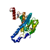
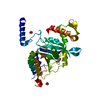
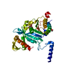
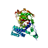
| Title | CRYSTAL STRUCTURE OF THE E121K SUBSTITUTION OF THE RECEIVER DOMAIN OF SINORHIZOBIUM MELILOTI DCTD | ||||||
 Components Components | C4-DICARBOXYLATE TRANSPORT TRANSCRIPTIONAL REGULATORY PROTEIN DCTD | ||||||
 Keywords Keywords | TRANSCRIPTION REGULATOR / two component receiver domain / sigma 54 / regulator of AAA+ ATPase | ||||||
| Function / homology |  Function and homology information Function and homology informationphosphorelay signal transduction system / sequence-specific DNA binding / regulation of DNA-templated transcription / DNA-templated transcription / ATP binding / cytoplasm Similarity search - Function | ||||||
| Biological species |  Sinorhizobium meliloti (bacteria) Sinorhizobium meliloti (bacteria) | ||||||
| Method |  X-RAY DIFFRACTION / MOLECULAR REPLACEMENT / Resolution: 2 Å X-RAY DIFFRACTION / MOLECULAR REPLACEMENT / Resolution: 2 Å | ||||||
 Authors Authors | Park, S. / Meyer, M. / Jones, A.D. / Yennawar, H.P. / Yennawar, N.H. / Nixon, B.T. | ||||||
 Citation Citation | Journal: FASEB J. / Year: 2002 Title: Two-component signaling in the AAA + ATPase DctD: binding Mg2+ and BeF3- selects between alternate dimeric states of the receiver domain Authors: Park, S. / Meyer, M. / Jones, A.D. / Yennawar, H.P. / Yennawar, N.H. / Nixon, B.T. #1: Journal: FASEB J. / Year: 2001Title: A dimeric two-component receiver domain inhibits the sigma54-dependent ATPase in DctD Authors: Meyer, M.G. / Park, S. / Zeringue, L. / Staley, M. / McKinstry, M. / Kaufman, R.I. / Zhang, H. / Yan, D. / Yennawar, N. / Yennawar, H. / Farber, G.K. / Nixon, B.T. | ||||||
| History |
|
- Structure visualization
| Structure viewer | Molecule: MolmilJmol/JSmol |
|---|
- Downloads & links
-Download
| PDBx/mmCIF format | 1l5z.cif.gz | 46.3 KB | Display | PDBx/mmCIF format |
|---|---|---|---|---|
| PDB format | pdb1l5z.ent.gz | 31.9 KB | Display | PDB format |
| PDBx/mmJSON format | 1l5z.json.gz | Tree view | PDBx/mmJSON format | |
| Others |  Other downloads Other downloads |
-Validation report
| Arichive directory | https://data.pdbj.org/pub/pdb/validation_reports/l5/1l5zftp://data.pdbj.org/pub/pdb/validation_reports/l5/1l5z | HTTPS FTP |
|---|
-Related structure data
| Related structure data |  1l5yC  1qkkS S: Starting model for refinement C: citing same article ( |
|---|---|
| Similar structure data |


-Links
 PDBj
PDBj

- Assembly
| Deposited unit | 
| ||||||||||||
|---|---|---|---|---|---|---|---|---|---|---|---|---|---|
| 1 | 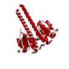
| ||||||||||||
| 2 | 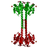
| ||||||||||||
| Unit cell |
| ||||||||||||
| Components on special symmetry positions |
| ||||||||||||
| Details | analytical ultracentrifuge data indicates solution form is a dimer; genetic and biochemical data indicates substitution destabilizes the dimer ~20-fold, leading to 'derepression' of the ATPase domain asymmetric unit is a monomer biological unit is a dimer - build with symmetry operation -X, -Y, Z |
-Components
| #1: Protein | Mass: 16821.326 Da / Num. of mol.: 1 / Fragment: RECEIVER DOMAIN, RESIDUES 2-143 / Mutation: E121K Source method: isolated from a genetically manipulated source Source: (gene. exp.) Sinorhizobium meliloti (bacteria) / Gene: dctD / Plasmid: pT143E121K / Production host: | ||
|---|---|---|---|
| #2: Chemical | ChemComp-SO4 /   Mass: 96.063 Da / Num. of mol.: 1 / Source method: obtained synthetically / Formula: SO4 Mass: 96.063 Da / Num. of mol.: 1 / Source method: obtained synthetically / Formula: SO4 | ||
| #3: Chemical | ChemComp-GOL /   Mass: 92.094 Da / Num. of mol.: 4 / Source method: obtained synthetically / Formula: C3H8O3 Mass: 92.094 Da / Num. of mol.: 4 / Source method: obtained synthetically / Formula: C3H8O3#4: Water | ChemComp-HOH / |  Mass: 18.015 Da / Num. of mol.: 144 / Source method: isolated from a natural source / Formula: H2O Mass: 18.015 Da / Num. of mol.: 144 / Source method: isolated from a natural source / Formula: H2O |
-Experimental details
-Experiment
| Experiment | Method: X-RAY DIFFRACTION / Number of used crystals: 1 |
|---|
- Sample preparation
| Crystal | Density Matthews: 4.17 Å3/Da / Density % sol: 70.47 % | |||||||||||||||||||||
|---|---|---|---|---|---|---|---|---|---|---|---|---|---|---|---|---|---|---|---|---|---|---|
| Crystal grow | Temperature: 298 K / Method: vapor diffusion, hanging drop / pH: 7.5 Details: HEPES, lithium sulfate, PEG 8000, pH 7.5, VAPOR DIFFUSION, HANGING DROP, temperature 298K | |||||||||||||||||||||
| Crystal grow | *PLUS pH: 5.6 / Method: vapor diffusion, hanging drop | |||||||||||||||||||||
| Components of the solutions | *PLUS
|
-Data collection
| Diffraction | Mean temperature: 100 K |
|---|---|
| Diffraction source | Source: ROTATING ANODE / Type: RIGAKU RU200 / Wavelength: 1.5418 Å |
| Detector | Type: RIGAKU RAXIS IV++ / Detector: IMAGE PLATE / Date: Sep 30, 2001 / Details: MSC Blue Confocal Optical System |
| Radiation | Monochromator: MSC Blue Confocal Optical System / Protocol: SINGLE WAVELENGTH / Monochromatic (M) / Laue (L): M / Scattering type: x-ray |
| Radiation wavelength | Wavelength: 1.5418 Å / Relative weight: 1 |
| Reflection | Resolution: 2→36.7 Å / Num. all: 19264 / Num. obs: 19264 / % possible obs: 98.6 % / Observed criterion σ(I): 2.2 / Redundancy: 3.04 % / Biso Wilson estimate: 16.6 Å2 / Rmerge(I) obs: 0.061 / Net I/σ(I): 7.1 |
| Reflection shell | Resolution: 2→2.07 Å / Redundancy: 1.61 % / Rmerge(I) obs: 0.222 / Mean I/σ(I) obs: 2.2 / Num. unique all: 3548 / % possible all: 97.5 |
| Reflection | *PLUS Highest resolution: 2 Å / % possible obs: 98.9 % / Num. measured all: 59135 |
| Reflection shell | *PLUS % possible obs: 98.1 % |
- Processing
| Software |
| ||||||||||||||||||||||||||||||||||||
|---|---|---|---|---|---|---|---|---|---|---|---|---|---|---|---|---|---|---|---|---|---|---|---|---|---|---|---|---|---|---|---|---|---|---|---|---|---|
| Refinement | Method to determine structure: MOLECULAR REPLACEMENT Starting model: PDB ENTRY 1QKK Resolution: 2→36.7 Å / Rfactor Rfree error: 0.005 / Data cutoff high absF: 215180.43 / Data cutoff low absF: 0 / Isotropic thermal model: RESTRAINED / Cross valid method: THROUGHOUT / σ(F): 0 / Stereochemistry target values: Engh & Huber
| ||||||||||||||||||||||||||||||||||||
| Solvent computation | Solvent model: FLAT MODEL / Bsol: 66.2093 Å2 / ksol: 0.385197 e/Å3 | ||||||||||||||||||||||||||||||||||||
| Displacement parameters | Biso mean: 22.4 Å2
| ||||||||||||||||||||||||||||||||||||
| Refine analyze |
| ||||||||||||||||||||||||||||||||||||
| Refinement step | Cycle: LAST / Resolution: 2→36.7 Å
| ||||||||||||||||||||||||||||||||||||
| Refine LS restraints |
| ||||||||||||||||||||||||||||||||||||
| LS refinement shell | Resolution: 2→2.13 Å / Rfactor Rfree error: 0.015 / Total num. of bins used: 6
| ||||||||||||||||||||||||||||||||||||
| Xplor file |
| ||||||||||||||||||||||||||||||||||||
| Refinement | *PLUS % reflection Rfree: 9.7 % | ||||||||||||||||||||||||||||||||||||
| Solvent computation | *PLUS | ||||||||||||||||||||||||||||||||||||
| Displacement parameters | *PLUS | ||||||||||||||||||||||||||||||||||||
| Refine LS restraints | *PLUS
|
