- PDB-1nri: Crystal Structure of Putative Phosphosugar Isomerase HI0754 from ... -
+
Open data
ID or keywords:
Loading...
-
Basic information
Entry
Database: PDB / ID: 1nri
Title
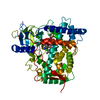
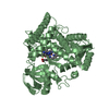
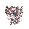
Crystal Structure of Putative Phosphosugar Isomerase HI0754 from Haemophilus influenzae
Components
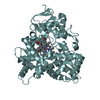
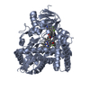
Hypothetical protein HI0754
Keywords
STRUCTURAL GENOMICS / UNKNOWN FUNCTION / Hypothetical Protein / Haemophilus influenzae / PSI / Protein Structure Initiative / Midwest Center for Structural Genomics / MCSG
Function / homology
Function and homology information
N-acetylmuramic acid 6-phosphate etherase / amino sugar catabolic process / N-acetylmuramic acid catabolic process / 1,6-anhydro-N-acetyl-beta-muramic acid catabolic process / ether hydrolase activity / carbon-oxygen lyase activity / peptidoglycan turnover / carbohydrate derivative binding Similarity search - Function
N-acetylmuramic acid 6-phosphate etherase MurQ / Glucokinase regulatory protein, conserved site / N-acetylmuramic acid 6-phosphate etherase/glucokinase regulatory protein / C-terminal lid domain of glucokinase regulatory protein / Glucokinase regulatory protein N-terminal SIS domain / Glucokinase regulatory protein family signature. / SIS domain / SIS domain / SIS domain profile. / SIS domain superfamily ...N-acetylmuramic acid 6-phosphate etherase MurQ / Glucokinase regulatory protein, conserved site / N-acetylmuramic acid 6-phosphate etherase/glucokinase regulatory protein / C-terminal lid domain of glucokinase regulatory protein / Glucokinase regulatory protein N-terminal SIS domain / Glucokinase regulatory protein family signature. / SIS domain / SIS domain / SIS domain profile. / SIS domain superfamily / Glucose-6-phosphate isomerase like protein; domain 1 / Rossmann fold / 3-Layer(aba) Sandwich / Alpha Beta Similarity search - Domain/homology
Resolution: 1.88→1.95 Å / Redundancy: 3.5 % / Rmerge(I) obs: 0.222 / Mean I/σ(I) obs: 5 / Num. unique all: 2198 / % possible all: 98.2
-
Processing
Software
Name
Version
Classification
CNS
1
refinement
d*TREK
datareduction
HKL-2000
datascaling
SHARP
phasing
Refinement
Method to determine structure: MAD / Resolution: 1.9→43.5 Å / Rfactor Rfree error: 0.005 / Isotropic thermal model: RESTRAINED / Cross valid method: THROUGHOUT / σ(F): 0 / Stereochemistry target values: Engh & Huber Details: The N-term 11 residues and the C-terminal 47 residues were not built due to poor electron density.
In the structure databanks used in Yorodumi, some data are registered as the other names, "COVID-19 virus" and "2019-nCoV". Here are the details of the virus and the list of structure data.
Jan 31, 2019. EMDB accession codes are about to change! (news from PDBe EMDB page)
EMDB accession codes are about to change! (news from PDBe EMDB page)
The allocation of 4 digits for EMDB accession codes will soon come to an end. Whilst these codes will remain in use, new EMDB accession codes will include an additional digit and will expand incrementally as the available range of codes is exhausted. The current 4-digit format prefixed with “EMD-” (i.e. EMD-XXXX) will advance to a 5-digit format (i.e. EMD-XXXXX), and so on. It is currently estimated that the 4-digit codes will be depleted around Spring 2019, at which point the 5-digit format will come into force.
The EM Navigator/Yorodumi systems omit the EMD- prefix.
Related info.:Q: What is EMD? / ID/Accession-code notation in Yorodumi/EM Navigator
Yorodumi is a browser for structure data from EMDB, PDB, SASBDB, etc.
This page is also the successor to EM Navigator detail page, and also detail information page/front-end page for Omokage search.
The word "yorodu" (or yorozu) is an old Japanese word meaning "ten thousand". "mi" (miru) is to see.
Related info.:EMDB / PDB / SASBDB / Comparison of 3 databanks / Yorodumi Search / Aug 31, 2016. New EM Navigator & Yorodumi / Yorodumi Papers / Jmol/JSmol / Function and homology information / Changes in new EM Navigator and Yorodumi
 Movie
Movie Controller

 Yorodumi
 Open data
 Basic information
 Components
 Keywords
 Function and homology information
 Haemophilus influenzae (bacteria)
 X-RAY DIFFRACTION /
  Authors
 Citation
 Structure visualization
 Downloads & links
 Other downloads


 PDBj
PDBj Assembly
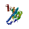

 Mass: 18.015 Da / Num. of mol.: 196 / Source method: isolated from a natural source / Formula: H2O
 Sample preparation
 Processing