 Movie
Movie Controller

[English] 日本語
 Yorodumi
- PDB-1qkk: Crystal structure of the receiver domain and linker region of Dct... -
+ Open data

- Basic information
| Entry | Database: PDB / ID: 1qkk | ||||||
|---|---|---|---|---|---|---|---|
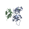
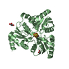
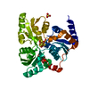
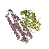
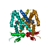
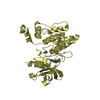
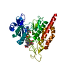
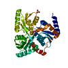
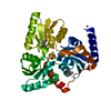
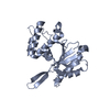
| Title | Crystal structure of the receiver domain and linker region of DctD from Sinorhizobium meliloti | ||||||
 Components Components | C4-DICARBOXYLATE TRANSPORT TRANSCRIPTIONAL REGULATORY PROTEIN | ||||||
 Keywords Keywords | TRANSCRIPTIONAL REGULATORY PROTEIN / RECEIVER DOMAIN / 2-COMPONENT SIGNAL TRANSDUCTION / SIGMA-54 DEPENDENT TRANSCRIPTIONAL ACTIVATOR / BACTERIAL ENHANCER BINDING PROTEIN / HIGH SOLVENT CONTENT CRYSTAL | ||||||
| Function / homology |  Function and homology information Function and homology informationphosphorelay signal transduction system / sequence-specific DNA binding / regulation of DNA-templated transcription / DNA-templated transcription / ATP binding / cytoplasm Similarity search - Function | ||||||
| Biological species |  SINORHIZOBIUM MELILOTI (bacteria) SINORHIZOBIUM MELILOTI (bacteria) | ||||||
| Method |  X-RAY DIFFRACTION / SYNCHROTRON / MAD / Resolution: 1.7 Å X-RAY DIFFRACTION / SYNCHROTRON / MAD / Resolution: 1.7 Å | ||||||
 Authors Authors | Meyer, M.G. / Park, S. / Zeringue, L. / Staley, M. / Mckinstry, M. / Kaufman, R.I. / Zhang, H. / Yan, D. / Yennawar, N. / Farber, G.K. / Nixon, B.T. | ||||||
 Citation Citation | Journal: Faseb J. / Year: 2001 Title: A dimeric two-component receiver domain inhibits the sigma54-dependent ATPase in DctD. Authors: Meyer, M.G. / Park, S. / Zeringue, L. / Staley, M. / McKinstry, M. / Kaufman, R.I. / Zhang, H. / Yan, D. / Yennawar, N. / Yennawar, H. / Farber, G.K. / Nixon, B.T. #1: Journal: Science / Year: 1987 Title: Crystallographic R Factor Refinement by Molecular Dynamics Authors: Brunger, A.T. / Kuriyan, J. / Karplus, M. | ||||||
| History |
|
- Structure visualization
| Structure viewer | Molecule: MolmilJmol/JSmol |
|---|
- Downloads & links
-Download
| PDBx/mmCIF format | 1qkk.cif.gz | 44.4 KB | Display | PDBx/mmCIF format |
|---|---|---|---|---|
| PDB format | pdb1qkk.ent.gz | 31.8 KB | Display | PDB format |
| PDBx/mmJSON format | 1qkk.json.gz | Tree view | PDBx/mmJSON format | |
| Others |  Other downloads Other downloads |
-Validation report
| Arichive directory | https://data.pdbj.org/pub/pdb/validation_reports/qk/1qkkftp://data.pdbj.org/pub/pdb/validation_reports/qk/1qkk | HTTPS FTP |
|---|
-Related structure data
| Similar structure data |
|---|
-Links
 PDBj
PDBj

- Assembly
| Deposited unit | 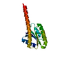
| ||||||||
|---|---|---|---|---|---|---|---|---|---|
| 1 | 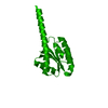
| ||||||||
| Unit cell |
| ||||||||
| Details | THE MOLECULE EXISTS AS A DIMER IN SOLUTION . THE DIMER ISCOMPRISED OF MONOMERS FROM DIFFERENT UNIT CELLS.DIMERIZATION SURFACES OCCUR AT THE FACES OF THE UNIT CELL"BOX". |
-Components
| #1: Protein | Mass: 16821.260 Da / Num. of mol.: 1 / Fragment: RESIDUES 2 TO 143, RECEIVER DOMAIN Source method: isolated from a genetically manipulated source Details: THE PROTEIN WAS EXPRESSED WITH THE C-TERMINAL HIS-TAG, KLAAALEHHHHHH. COORDINATES ARE SUBMITTED ONLY FOR THE MONOMER, WHICH COMPRISES THE COMPLETE ASYMMETRIC UNIT. Source: (gene. exp.) SINORHIZOBIUM MELILOTI (bacteria) / Strain: N.A. 1021 / Plasmid: PET21A / Production host: |
|---|---|
| #2: Water | ChemComp-HOH /  Mass: 18.015 Da / Num. of mol.: 204 / Source method: isolated from a natural source / Formula: H2O Mass: 18.015 Da / Num. of mol.: 204 / Source method: isolated from a natural source / Formula: H2O |
| Compound details | CHAIN A: RESIDUES 144 TO 156 ARE A HIS TAG |
| Source details | THE PROTEIN IS FROM N.A. STRAIN=1021 REFERENCE: JIANG J., GU B., ALBRIGHT L.M., NIXON B.T., ...THE PROTEIN IS FROM N.A. STRAIN=1021 REFERENCE: JIANG J., GU B., ALBRIGHT L.M., NIXON B.T., CONSERVATI |
-Experimental details
-Experiment
| Experiment | Method: X-RAY DIFFRACTION / Number of used crystals: 2 |
|---|
- Sample preparation
| Crystal | Density Matthews: 4.3 Å3/Da / Density % sol: 71 % Description: DATA WERE COLLECTED AT BEAMLINE 17-ID (OR 17-BM) IN THE FACILITIES OF THEINDUSTRIAL MACROMOLECULAR CRYSTALLOGRAPHY ASSOCIATION COLLABORATIVE ACCESS TEAM (IMCA-CAT) AT THE ADVANCED PHOTON ...Description: DATA WERE COLLECTED AT BEAMLINE 17-ID (OR 17-BM) IN THE FACILITIES OF THEINDUSTRIAL MACROMOLECULAR CRYSTALLOGRAPHY ASSOCIATION COLLABORATIVE ACCESS TEAM (IMCA-CAT) AT THE ADVANCED PHOTON SOURCE.THESE FACILITIES ARE SUPPORTED BY THE COMPANIES OF THE INDUSTRIAL MACROMOLECULAR CRYSTALLOGRAPHY ASSOCIATION THROUGH A CONTRACT WITH ILLINOIS INSTITUTE OF TECHNOLOGY (IIT), EXECUTED THROUGH THE IIT'S CENTER FOR SYNCHROTRON RADIATION RESEARCH AND INSTRUMENTATION. | ||||||||||||||||||||||||
|---|---|---|---|---|---|---|---|---|---|---|---|---|---|---|---|---|---|---|---|---|---|---|---|---|---|
| Crystal grow | pH: 5.6 Details: 50 MM NA SUCCINATE PH 5.6, 70 MM AMMONIUM PHOSPHATE MONOBASIC, 1 MM DTT | ||||||||||||||||||||||||
| Crystal grow | *PLUS Temperature: 294 K / Method: vapor diffusion, hanging drop | ||||||||||||||||||||||||
| Components of the solutions | *PLUS
|
-Data collection
| Diffraction | Mean temperature: 100 K | ||||||||||||
|---|---|---|---|---|---|---|---|---|---|---|---|---|---|
| Diffraction source | Source: SYNCHROTRON / Site: APS  / Beamline: 17-ID / Wavelength: 0.98333, 0.98100, 0.98076 / Beamline: 17-ID / Wavelength: 0.98333, 0.98100, 0.98076 | ||||||||||||
| Detector | Type: MARRESEARCH / Detector: CCD / Date: Nov 27, 1998 / Details: NO FOCUSSING MIRROR | ||||||||||||
| Radiation | Monochromator: CRYOGENICALLY COOLED SI (111) MONOCHROMATOR / Protocol: MAD / Monochromatic (M) / Laue (L): M / Scattering type: x-ray | ||||||||||||
| Radiation wavelength |
| ||||||||||||
| Reflection | Resolution: 1.7→20 Å / Num. obs: 61207 / % possible obs: 99.6 % / Observed criterion σ(I): 2 / Redundancy: 8 % / Biso Wilson estimate: 20 Å2 / Rsym value: 0.062 / Net I/σ(I): 34 | ||||||||||||
| Reflection shell | Resolution: 1.7→1.76 Å / Redundancy: 6.5 % / Mean I/σ(I) obs: 2.2 / Rsym value: 0.324 / % possible all: 100 | ||||||||||||
| Reflection | *PLUS Num. obs: 32439 / % possible obs: 86.1 % / Rmerge(I) obs: 0.059 |
- Processing
| Software |
| ||||||||||||||||||||||||||||||||||||||||||||||||||||||||||||||||||||||||||||||||
|---|---|---|---|---|---|---|---|---|---|---|---|---|---|---|---|---|---|---|---|---|---|---|---|---|---|---|---|---|---|---|---|---|---|---|---|---|---|---|---|---|---|---|---|---|---|---|---|---|---|---|---|---|---|---|---|---|---|---|---|---|---|---|---|---|---|---|---|---|---|---|---|---|---|---|---|---|---|---|---|---|---|
| Refinement | Method to determine structure: MAD / Resolution: 1.7→19.42 Å / Rfactor Rfree error: 0.005 / Data cutoff high absF: 1822253.53 / Data cutoff low absF: 0 / Isotropic thermal model: RESTRAINED / Cross valid method: THROUGHOUT / σ(F): 0
| ||||||||||||||||||||||||||||||||||||||||||||||||||||||||||||||||||||||||||||||||
| Solvent computation | Solvent model: FLAT MODEL / Bsol: 64.1063 Å2 / ksol: 0.415432 e/Å3 | ||||||||||||||||||||||||||||||||||||||||||||||||||||||||||||||||||||||||||||||||
| Displacement parameters | Biso mean: 27 Å2
| ||||||||||||||||||||||||||||||||||||||||||||||||||||||||||||||||||||||||||||||||
| Refine analyze |
| ||||||||||||||||||||||||||||||||||||||||||||||||||||||||||||||||||||||||||||||||
| Refinement step | Cycle: LAST / Resolution: 1.7→19.42 Å
| ||||||||||||||||||||||||||||||||||||||||||||||||||||||||||||||||||||||||||||||||
| Refine LS restraints |
| ||||||||||||||||||||||||||||||||||||||||||||||||||||||||||||||||||||||||||||||||
| LS refinement shell | Resolution: 1.7→1.81 Å / Rfactor Rfree error: 0.013 / Total num. of bins used: 6
| ||||||||||||||||||||||||||||||||||||||||||||||||||||||||||||||||||||||||||||||||
| Xplor file |
| ||||||||||||||||||||||||||||||||||||||||||||||||||||||||||||||||||||||||||||||||
| Software | *PLUS Name: CNS / Version: 0.9 / Classification: refinement | ||||||||||||||||||||||||||||||||||||||||||||||||||||||||||||||||||||||||||||||||
| Refinement | *PLUS Rfactor Rfree: 0.231 / Rfactor Rwork: 0.201 | ||||||||||||||||||||||||||||||||||||||||||||||||||||||||||||||||||||||||||||||||
| Solvent computation | *PLUS | ||||||||||||||||||||||||||||||||||||||||||||||||||||||||||||||||||||||||||||||||
| Displacement parameters | *PLUS | ||||||||||||||||||||||||||||||||||||||||||||||||||||||||||||||||||||||||||||||||
| Refine LS restraints | *PLUS
|
