 Movie
Movie Controller

[English] 日本語
 Yorodumi
- PDB-1l5x: The 2.0-Angstrom resolution crystal structure of a survival prote... -
+ Open data

- Basic information
| Entry | Database: PDB / ID: 1l5x | ||||||
|---|---|---|---|---|---|---|---|
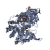
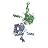
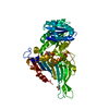
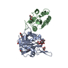
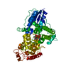
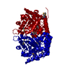
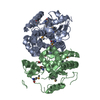
| Title | The 2.0-Angstrom resolution crystal structure of a survival protein E (SurE) homolog from Pyrobaculum aerophilum | ||||||
 Components Components | Survival protein E | ||||||
 Keywords Keywords | STRUCTURAL GENOMICS / UNKNOWN FUNCTION / putative acid phosphatase / mixed alpha/beta protein / N-terminal Rossmann-fold like / novel C-terminal domain with beta-hairpin extensions | ||||||
| Function / homology |  Function and homology information Function and homology information5'-deoxynucleotidase activity / : / 5'-nucleotidase / nucleotide binding / metal ion binding / cytoplasm Similarity search - Function | ||||||
| Biological species |   Pyrobaculum aerophilum (archaea) Pyrobaculum aerophilum (archaea) | ||||||
| Method |  X-RAY DIFFRACTION / SYNCHROTRON / MAD / Resolution: 2 Å X-RAY DIFFRACTION / SYNCHROTRON / MAD / Resolution: 2 Å | ||||||
 Authors Authors | Mura, C. / Katz, J.E. / Clarke, S.G. / Eisenberg, D. | ||||||
 Citation Citation | Journal: J.Mol.Biol. / Year: 2003 Title: Structure and Function of an Archaeal Homolog of Survival Protein E (SurE-alpha): An Acid Phosphatase with Purine Nucleotide Specificity Authors: Mura, C. / Katz, J.E. / Clarke, S.G. / Eisenberg, D. #1: Journal: Nat.Struct.Biol. / Year: 2001Title: Crystal Structure and Functional Analysis of the SurE Protein Identify a Novel Phosphatase Family Authors: Lee, J.Y. / Kwak, J.E. / Moon, J. / Eom, S.H. / Liong, E.C. / Pedelacq, J.-D. / Berendzen, J. / Suh, S.W. #2: Journal: Structure / Year: 2001Title: Structure of Thermotoga maritima Stationary Phase Survival Protein SurE: A Novel Acid Phosphatase Authors: Zhang, R.-G. / Skarina, T. / Katz, J.E. / Beasley, S. / Khachatryan, A. / Vyas, S. / Arrowsmith, C.H. / Clarke, S. / Edwards, A. / Joachimiak, A. | ||||||
| History |
| ||||||
| Remark 400 | COMPOUND THE FIRST STRUCTURE OF AN ARCHAEAL SURE, WITH STRONG SIMILARITY TO THE PROKARYOTIC SURE ...COMPOUND THE FIRST STRUCTURE OF AN ARCHAEAL SURE, WITH STRONG SIMILARITY TO THE PROKARYOTIC SURE STRUCTURE FROM THERMATOGA MARITIMA (SEE RELATED ENTRIES). | ||||||
| Remark 999 | SEQUENCE THE ADDITIONAL RESIDUES FROM SER 267 ONWARDS ARE CLONING ARTIFACTS (HIS-TAG AND LINKER). |
- Structure visualization
| Structure viewer | Molecule: MolmilJmol/JSmol |
|---|
- Downloads & links
-Download
| PDBx/mmCIF format | 1l5x.cif.gz | 125.4 KB | Display | PDBx/mmCIF format |
|---|---|---|---|---|
| PDB format | pdb1l5x.ent.gz | 98.5 KB | Display | PDB format |
| PDBx/mmJSON format | 1l5x.json.gz | Tree view | PDBx/mmJSON format | |
| Others |  Other downloads Other downloads |
-Validation report
| Arichive directory | https://data.pdbj.org/pub/pdb/validation_reports/l5/1l5xftp://data.pdbj.org/pub/pdb/validation_reports/l5/1l5x | HTTPS FTP |
|---|
-Related structure data
| Related structure data | |
|---|---|
| Similar structure data |


-Links
 PDBj
PDBj

- Assembly
| Deposited unit | 
| ||||||||
|---|---|---|---|---|---|---|---|---|---|
| 1 |
| ||||||||
| 2 | 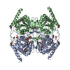
| ||||||||
| Unit cell |
| ||||||||
| Details | The biological assembly is thought to be the dimer found in the asymmetric unit of this deposition. |
-Components
| #1: Protein | Mass: 30768.275 Da / Num. of mol.: 2 Source method: isolated from a genetically manipulated source Source: (gene. exp.) Pyrobaculum aerophilum (archaea) / Gene: PAE2908 / Plasmid: pET-22b(+) / Species (production host): Escherichia coli / Production host:  #2: Chemical | ChemComp-GOL /   Mass: 92.094 Da / Num. of mol.: 7 / Source method: obtained synthetically / Formula: C3H8O3 Mass: 92.094 Da / Num. of mol.: 7 / Source method: obtained synthetically / Formula: C3H8O3#3: Chemical |   Mass: 60.052 Da / Num. of mol.: 2 / Source method: obtained synthetically / Formula: C2H4O2 Mass: 60.052 Da / Num. of mol.: 2 / Source method: obtained synthetically / Formula: C2H4O2#4: Water | ChemComp-HOH / |  Mass: 18.015 Da / Num. of mol.: 287 / Source method: isolated from a natural source / Formula: H2O Mass: 18.015 Da / Num. of mol.: 287 / Source method: isolated from a natural source / Formula: H2OHas protein modification | Y | |
|---|
-Experimental details
-Experiment
| Experiment | Method: X-RAY DIFFRACTION / Number of used crystals: 1 |
|---|
- Sample preparation
| Crystal | Density Matthews: 2.5 Å3/Da / Density % sol: 50.71 % | ||||||||||||||||||||||||||||||||||||
|---|---|---|---|---|---|---|---|---|---|---|---|---|---|---|---|---|---|---|---|---|---|---|---|---|---|---|---|---|---|---|---|---|---|---|---|---|---|
| Crystal grow | Temperature: 293 K / Method: vapor diffusion, hanging drop / pH: 8.55 Details: PEG 4000, sodium acetate, glycerol, Tris buffer, pH 8.55, VAPOR DIFFUSION, HANGING DROP, temperature 293.0K | ||||||||||||||||||||||||||||||||||||
| Crystal grow | *PLUS Temperature: 19.8 ℃ | ||||||||||||||||||||||||||||||||||||
| Components of the solutions | *PLUS
|
-Data collection
| Diffraction | Mean temperature: 105 K | ||||||||||||
|---|---|---|---|---|---|---|---|---|---|---|---|---|---|
| Diffraction source | Source: SYNCHROTRON / Site: ALS  / Beamline: 5.0.2 / Wavelength: 0.97870, 0.97860, 0.96485 / Beamline: 5.0.2 / Wavelength: 0.97870, 0.97860, 0.96485 | ||||||||||||
| Detector | Type: ADSC QUANTUM 4 / Detector: CCD / Date: Jan 18, 2002 | ||||||||||||
| Radiation | Monochromator: double crystal monochromator / Protocol: MAD / Monochromatic (M) / Laue (L): M / Scattering type: x-ray | ||||||||||||
| Radiation wavelength |
| ||||||||||||
| Reflection | Resolution: 2→100 Å / Num. all: 42129 / Num. obs: 42129 / % possible obs: 99.9 % / Observed criterion σ(F): -3 / Observed criterion σ(I): -3 / Redundancy: 7.1 % / Biso Wilson estimate: 18.1 Å2 / Rmerge(I) obs: 0.099 / Rsym value: 0.099 / Net I/σ(I): 17.4 | ||||||||||||
| Reflection shell | Resolution: 2→2.07 Å / Redundancy: 7 % / Rmerge(I) obs: 0.781 / Mean I/σ(I) obs: 2.5 / Rsym value: 0.781 / % possible all: 100 | ||||||||||||
| Reflection | *PLUS Lowest resolution: 100 Å / Num. measured all: 297069 | ||||||||||||
| Reflection shell | *PLUS % possible obs: 100 % |
- Processing
| Software |
| ||||||||||||||||||||||||||||||||||||
|---|---|---|---|---|---|---|---|---|---|---|---|---|---|---|---|---|---|---|---|---|---|---|---|---|---|---|---|---|---|---|---|---|---|---|---|---|---|
| Refinement | Method to determine structure: MAD / Resolution: 2→19.59 Å / Rfactor Rfree error: 0.005 / Data cutoff high absF: 195285.23 / Data cutoff low absF: 0 / Isotropic thermal model: RESTRAINED / Cross valid method: THROUGHOUT / σ(F): 0 / Stereochemistry target values: Engh & Huber
| ||||||||||||||||||||||||||||||||||||
| Solvent computation | Solvent model: FLAT MODEL / Bsol: 61.4539 Å2 / ksol: 0.393386 e/Å3 | ||||||||||||||||||||||||||||||||||||
| Displacement parameters | Biso mean: 37.3 Å2
| ||||||||||||||||||||||||||||||||||||
| Refine analyze |
| ||||||||||||||||||||||||||||||||||||
| Refinement step | Cycle: LAST / Resolution: 2→19.59 Å
| ||||||||||||||||||||||||||||||||||||
| Refine LS restraints |
| ||||||||||||||||||||||||||||||||||||
| LS refinement shell | Resolution: 2→2.13 Å / Rfactor Rfree error: 0.016 / Total num. of bins used: 6
| ||||||||||||||||||||||||||||||||||||
| Xplor file |
| ||||||||||||||||||||||||||||||||||||
| Refinement | *PLUS Highest resolution: 2 Å / Lowest resolution: 20 Å / % reflection Rfree: 4.6 % | ||||||||||||||||||||||||||||||||||||
| Solvent computation | *PLUS | ||||||||||||||||||||||||||||||||||||
| Displacement parameters | *PLUS | ||||||||||||||||||||||||||||||||||||
| Refine LS restraints | *PLUS
|
