 Movie
Movie Controller


+ Open data

- Basic information
| Entry | Database: PDB / ID: 1l5a | ||||||
|---|---|---|---|---|---|---|---|
| Title | Crystal Structure of VibH, an NRPS Condensation Enzyme | ||||||
 Components Components | amide synthase | ||||||
 Keywords Keywords | BIOSYNTHETIC PROTEIN / nonribosomal peptide synthetase / NRPS condensation domain / amide synthase / vibriobactin | ||||||
| Function / homology |  Function and homology information Function and homology informationacid-ammonia (or amide) ligase activity / siderophore biosynthetic process Similarity search - Function | ||||||
| Biological species |   Vibrio cholerae (bacteria) Vibrio cholerae (bacteria) | ||||||
| Method |  X-RAY DIFFRACTION / SYNCHROTRON / MAD / Resolution: 2.55 Å X-RAY DIFFRACTION / SYNCHROTRON / MAD / Resolution: 2.55 Å | ||||||
 Authors Authors | Keating, T.A. / Marshall, C.G. / Walsh, C.T. / Keating, A.E. | ||||||
 Citation Citation | Journal: Nat.Struct.Biol. / Year: 2002 Title: The structure of VibH represents nonribosomal peptide synthetase condensation, cyclization and epimerization domains. Authors: Keating, T.A. / Marshall, C.G. / Walsh, C.T. / Keating, A.E. | ||||||
| History |
|
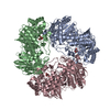
- Structure visualization
| Structure viewer | Molecule: MolmilJmol/JSmol |
|---|
- Downloads & links
-Download
| PDBx/mmCIF format | 1l5a.cif.gz | 257.6 KB | Display | PDBx/mmCIF format |
|---|---|---|---|---|
| PDB format | pdb1l5a.ent.gz | 210.8 KB | Display | PDB format |
| PDBx/mmJSON format | 1l5a.json.gz | Tree view | PDBx/mmJSON format | |
| Others |  Other downloads Other downloads |
-Validation report
| Summary document | 1l5a_validation.pdf.gz | 445.8 KB | Display | wwPDB validaton report |
|---|---|---|---|---|
| Full document | 1l5a_full_validation.pdf.gz | 475.1 KB | Display | |
| Data in XML | 1l5a_validation.xml.gz | 48 KB | Display | |
| Data in CIF | 1l5a_validation.cif.gz | 66.8 KB | Display | |
| Arichive directory | https://data.pdbj.org/pub/pdb/validation_reports/l5/1l5aftp://data.pdbj.org/pub/pdb/validation_reports/l5/1l5a | HTTPS FTP |
-Related structure data
| Similar structure data |
|---|


-Links
 PDBj
PDBj

- Assembly
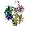
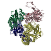
| Deposited unit | 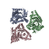
| ||||||||
|---|---|---|---|---|---|---|---|---|---|
| 1 |
| ||||||||
| Unit cell |
|
-Components
| #1: Protein | Mass: 49795.074 Da / Num. of mol.: 3 Source method: isolated from a genetically manipulated source Source: (gene. exp.) Vibrio cholerae (bacteria) / Gene: vibH / Plasmid: pET29b / Species (production host): Escherichia coli / Production host: #2: Water | ChemComp-HOH / |  Mass: 18.015 Da / Num. of mol.: 194 / Source method: isolated from a natural source / Formula: H2O Mass: 18.015 Da / Num. of mol.: 194 / Source method: isolated from a natural source / Formula: H2O |
|---|
-Experimental details
-Experiment
| Experiment | Method: X-RAY DIFFRACTION / Number of used crystals: 2 |
|---|
- Sample preparation
| Crystal | Density Matthews: 4 Å3/Da / Density % sol: 69 % | ||||||||||||||||||||||||||||||
|---|---|---|---|---|---|---|---|---|---|---|---|---|---|---|---|---|---|---|---|---|---|---|---|---|---|---|---|---|---|---|---|
| Crystal grow | Temperature: 293 K / Method: vapor diffusion, hanging drop / pH: 8.5 Details: PEG 8000, Tris, magnesium formate, pH 8.5, VAPOR DIFFUSION, HANGING DROP, temperature 293K | ||||||||||||||||||||||||||||||
| Crystal grow | *PLUS Temperature: 20 ℃ / Details: used microseeding | ||||||||||||||||||||||||||||||
| Components of the solutions | *PLUS
|
-Data collection
| Diffraction |
| |||||||||||||||
|---|---|---|---|---|---|---|---|---|---|---|---|---|---|---|---|---|
| Diffraction source |
| |||||||||||||||
| Detector | Type: BRANDEIS - B4 / Detector: CCD / Date: May 21, 2001 | |||||||||||||||
| Radiation | Monochromator: double crystal, torroidal mirror / Protocol: MAD / Monochromatic (M) / Laue (L): M / Scattering type: x-ray | |||||||||||||||
| Radiation wavelength |
| |||||||||||||||
| Reflection | Resolution: 2.55→35 Å / Num. all: 78790 / Num. obs: 73969 / % possible obs: 93.9 % / Observed criterion σ(F): 0 / Observed criterion σ(I): -3 / Redundancy: 2.6 % / Biso Wilson estimate: 55.5 Å2 / Rsym value: 0.046 / Net I/σ(I): 19.1 | |||||||||||||||
| Reflection shell | Resolution: 2.55→2.64 Å / Num. unique all: 4581 / Rsym value: 0.321 / % possible all: 58.5 | |||||||||||||||
| Reflection | *PLUS Lowest resolution: 35 Å / Rmerge(I) obs: 0.046 | |||||||||||||||
| Reflection shell | *PLUS Rmerge(I) obs: 0.32 |

- Processing
| Software |
| |||||||||||||||||||||||||
|---|---|---|---|---|---|---|---|---|---|---|---|---|---|---|---|---|---|---|---|---|---|---|---|---|---|---|
| Refinement | Method to determine structure: MAD / Resolution: 2.55→34.14 Å / Rfactor Rfree error: 0.003 / Data cutoff high absF: 2053567.31 / Data cutoff high rms absF: 2053567.31 / Isotropic thermal model: restrained / Cross valid method: THROUGHOUT / σ(F): 0 / Stereochemistry target values: Engh & Huber
| |||||||||||||||||||||||||
| Solvent computation | Solvent model: FLAT MODEL / Bsol: 33.7813 Å2 / ksol: 0.302009 e/Å3 | |||||||||||||||||||||||||
| Displacement parameters | Biso mean: 47.6 Å2
| |||||||||||||||||||||||||
| Refine analyze |
| |||||||||||||||||||||||||
| Refinement step | Cycle: LAST / Resolution: 2.55→34.14 Å
| |||||||||||||||||||||||||
| Refine LS restraints |
| |||||||||||||||||||||||||
| LS refinement shell | Resolution: 2.55→2.71 Å / Rfactor Rfree error: 0.012 / Total num. of bins used: 6
| |||||||||||||||||||||||||
| Refinement | *PLUS % reflection Rfree: 10 % / Rfactor obs: 0.214 / Rfactor Rfree: 0.259 / Rfactor Rwork: 0.214 | |||||||||||||||||||||||||
| Solvent computation | *PLUS | |||||||||||||||||||||||||
| Displacement parameters | *PLUS | |||||||||||||||||||||||||
| Refine LS restraints | *PLUS
| |||||||||||||||||||||||||
| LS refinement shell | *PLUS Rfactor Rfree: 0.377 / Rfactor Rwork: 0.314 |
