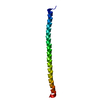
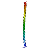
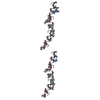
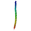
登録情報 データベース : PDB / ID : 1l2pタイトル ATP Synthase b Subunit Dimerization Domain ATP Synthase B Chain キーワード / 機能・相同性 分子機能 ドメイン・相同性 構成要素
/ / / / / / / / / / / / / 生物種 Escherichia coli (大腸菌)手法 / / / 解像度 : 1.55 Å データ登録者 Del Rizzo, P.A. / Dunn, S.D. / Bi, Y. / Shilton, B.H. ジャーナル : Biochemistry / 年 : 2002タイトル : The "second stalk" of Escherichia coli ATP synthase: structure of the isolated dimerization domain.著者 : Del Rizzo, P.A. / Bi, Y. / Dunn, S.D. / Shilton, B.H. 履歴 登録 2002年2月22日 登録サイト / 処理サイト 改定 1.0 2002年6月5日 Provider / タイプ 改定 1.1 2008年4月28日 Group 改定 1.2 2011年7月13日 Group 改定 1.3 2018年1月31日 Group / カテゴリ / Item 改定 1.4 2024年2月14日 Group / Data collection / Database referencesカテゴリ chem_comp_atom / chem_comp_bond ... chem_comp_atom / chem_comp_bond / database_2 / pdbx_database_remark Item / _database_2.pdbx_database_accession / _pdbx_database_remark.text
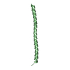
すべて表示 表示を減らす Remark 300 BIOMOLECULE: 1 THIS ENTRY CONTAINS THE CRYSTALLOGRAPHIC ASYMMETRIC UNIT WHICH CONSISTS OF 1 ... BIOMOLECULE: 1 THIS ENTRY CONTAINS THE CRYSTALLOGRAPHIC ASYMMETRIC UNIT WHICH CONSISTS OF 1 CHAIN(S). The biological unit is a dimer. However, the biological dimer is not contained in the crystal. The dimer model cannot be generated by a simple coordinate transformation of the crystal structure. The biological dimer can, however, be modelled based on the surface properties of the crystal structure, and on solution small-angle X-ray scattering data.
 ムービー
ムービー コントローラー


 データを開く
 基本情報
基本情報 要素
要素 キーワード
キーワード 機能・相同性情報
 X線回折 /
  データ登録者
 引用
引用 構造の表示
構造の表示 ダウンロードとリンク
 その他のダウンロード
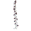

 PDBj
PDBj
 集合体
集合体
 分子量: 18.015 Da / 分子数: 54 / 由来タイプ: 天然 / 式: H2O
 試料調製
試料調製 / ビームライン: F1 / 波長: 0.99203 Å
 解析
解析