 Movie
Movie Controller


+ Open data

- Basic information
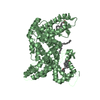
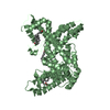
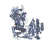
| Entry | Database: PDB / ID: 1kw2 | ||||||
|---|---|---|---|---|---|---|---|
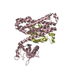
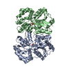
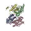
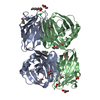
| Title | CRYSTAL STRUCTURE OF UNCOMPLEXED VITAMIN D-BINDING PROTEIN | ||||||
 Components Components | Vitamin D-binding protein | ||||||
 Keywords Keywords | TRANSPORT PROTEIN / DBP / vitamin D-binding protein / Actin scavenger system / Actin-binding protein | ||||||
| Function / homology |  Function and homology information Function and homology informationvitamin transmembrane transporter activity / calcidiol binding / vitamin transport / Vitamin D (calciferol) metabolism / vitamin D metabolic process / vitamin D binding / lysosomal lumen / response to nutrient levels / actin binding / blood microparticle ...vitamin transmembrane transporter activity / calcidiol binding / vitamin transport / Vitamin D (calciferol) metabolism / vitamin D metabolic process / vitamin D binding / lysosomal lumen / response to nutrient levels / actin binding / blood microparticle / : / extracellular exosome / extracellular region / cytosol Similarity search - Function | ||||||
| Biological species |  Homo sapiens (human) Homo sapiens (human) | ||||||
| Method |  X-RAY DIFFRACTION / SYNCHROTRON / MOLECULAR REPLACEMENT / Resolution: 2.15 Å X-RAY DIFFRACTION / SYNCHROTRON / MOLECULAR REPLACEMENT / Resolution: 2.15 Å | ||||||
 Authors Authors | Otterbein, L.R. / Dominguez, R. | ||||||
 Citation Citation | Journal: Proc.Natl.Acad.Sci.USA / Year: 2002 Title: Crystal structures of the vitamin D-binding protein and its complex with actin: structural basis of the actin-scavenger system. Authors: Otterbein, L.R. / Cosio, C. / Graceffa, P. / Dominguez, R. | ||||||
| History |
|
- Structure visualization
| Structure viewer | Molecule: MolmilJmol/JSmol |
|---|
- Downloads & links
-Download
| PDBx/mmCIF format | 1kw2.cif.gz | 189.2 KB | Display | PDBx/mmCIF format |
|---|---|---|---|---|
| PDB format | pdb1kw2.ent.gz | 151 KB | Display | PDB format |
| PDBx/mmJSON format | 1kw2.json.gz | Tree view | PDBx/mmJSON format | |
| Others |  Other downloads Other downloads |
-Validation report
| Arichive directory | https://data.pdbj.org/pub/pdb/validation_reports/kw/1kw2ftp://data.pdbj.org/pub/pdb/validation_reports/kw/1kw2 | HTTPS FTP |
|---|
-Related structure data
| Related structure data | 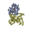 1kxpSC S: Starting model for refinement C: citing same article ( |
|---|---|
| Similar structure data |


-Links
 PDBj
PDBj


- Assembly
| Deposited unit | 
| ||||||||
|---|---|---|---|---|---|---|---|---|---|
| 1 | 
| ||||||||
| 2 | 
| ||||||||
| Unit cell |
| ||||||||
| Details | Two molecules are present in the asymetric unit. Molecule A is the best one. |
-Components
| #1: Protein | Mass: 51305.367 Da / Num. of mol.: 2 / Source method: isolated from a natural source / Details: serum / Source: (natural) Homo sapiens (human) / References: UniProt: P02774#2: Water | ChemComp-HOH / |  Mass: 18.015 Da / Num. of mol.: 333 / Source method: isolated from a natural source / Formula: H2O Mass: 18.015 Da / Num. of mol.: 333 / Source method: isolated from a natural source / Formula: H2OHas protein modification | Y | |
|---|
-Experimental details
-Experiment
| Experiment | Method: X-RAY DIFFRACTION / Number of used crystals: 1 |
|---|
- Sample preparation
| Crystal | Density Matthews: 3.2 Å3/Da / Density % sol: 61.8 % | ||||||||||||||||||||||||
|---|---|---|---|---|---|---|---|---|---|---|---|---|---|---|---|---|---|---|---|---|---|---|---|---|---|
| Crystal grow | Temperature: 293 K / Method: vapor diffusion, hanging drop / pH: 4.6 Details: PEG 200, 28%, Sodium Acetate, 0.1M, pH 4.6, VAPOR DIFFUSION, HANGING DROP, temperature 293K | ||||||||||||||||||||||||
| Crystal grow | *PLUS Temperature: 20 ℃ | ||||||||||||||||||||||||
| Components of the solutions | *PLUS
|
-Data collection
| Diffraction | Mean temperature: 100 K |
|---|---|
| Diffraction source | Source: SYNCHROTRON / Site: APS  / Beamline: 14-BM-C / Wavelength: 1 Å / Beamline: 14-BM-C / Wavelength: 1 Å |
| Detector | Type: ADSC QUANTUM 4 / Detector: CCD / Date: Dec 9, 2001 / Details: BENT CYLINDRICAL Si-MIRROR (RH coating) |
| Radiation | Monochromator: Si (III)DOUBLE CRYSTAL,L=1 / Protocol: SINGLE WAVELENGTH / Monochromatic (M) / Laue (L): M / Scattering type: x-ray |
| Radiation wavelength | Wavelength: 1 Å / Relative weight: 1 |
| Reflection | Resolution: 2.15→60 Å / Num. all: 67333 / Num. obs: 67333 / % possible obs: 94.9 % / Redundancy: 15.5 % / Biso Wilson estimate: 44 Å2 / Rmerge(I) obs: 0.059 / Net I/σ(I): 27.7 |
| Reflection shell | Resolution: 2.15→2.32 Å / Redundancy: 8.1 % / Rmerge(I) obs: 0.214 / Mean I/σ(I) obs: 10.2 / Num. unique all: 16595 / % possible all: 81.2 |
| Reflection | *PLUS Lowest resolution: 60 Å / Num. obs: 67175 / Num. measured all: 1041011 |
| Reflection shell | *PLUS % possible obs: 81.2 % |
- Processing
| Software |
| |||||||||||||||||||||||||
|---|---|---|---|---|---|---|---|---|---|---|---|---|---|---|---|---|---|---|---|---|---|---|---|---|---|---|
| Refinement | Method to determine structure: MOLECULAR REPLACEMENT Starting model: VITAMIN D-BINDING PROTEIN COMPLEXED WITH ACTIN PDB 1KXP Resolution: 2.15→60 Å / Cross valid method: THROUGHOUT / σ(F): 0 / σ(I): 0
| |||||||||||||||||||||||||
| Refine analyze |
| |||||||||||||||||||||||||
| Refinement step | Cycle: LAST / Resolution: 2.15→60 Å
| |||||||||||||||||||||||||
| Refine LS restraints |
| |||||||||||||||||||||||||
| LS refinement shell | Resolution: 2.15→2.32 Å
| |||||||||||||||||||||||||
| Xplor file | Serial no: 1 / Param file: PROTEIN_REP.PARM | |||||||||||||||||||||||||
| Refinement | *PLUS % reflection Rfree: 5 % | |||||||||||||||||||||||||
| Solvent computation | *PLUS | |||||||||||||||||||||||||
| Displacement parameters | *PLUS | |||||||||||||||||||||||||
| Refine LS restraints | *PLUS
| |||||||||||||||||||||||||
| LS refinement shell | *PLUS Rfactor Rwork: 0.251 |
