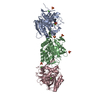
 Movie
Movie Controller

[English] 日本語
 Yorodumi
- PDB-1kvk: The Structure of Binary complex between a Mammalian Mevalonate Ki... -
+ Open data

- Basic information
| Entry | Database: PDB / ID: 1kvk | ||||||
|---|---|---|---|---|---|---|---|
| Title | The Structure of Binary complex between a Mammalian Mevalonate Kinase and ATP: Insights into the Reaction Mechanism and Human Inherited Disease | ||||||
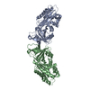
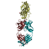
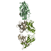
 Components Components | mevalonate kinase | ||||||
 Keywords Keywords | TRANSFERASE / RMK / ATP / GHMP | ||||||
| Function / homology |  Function and homology information Function and homology informationLanosterol biosynthesis / zymosterol biosynthetic process / mevalonate kinase / mevalonate kinase activity / : / : / farnesyl diphosphate biosynthetic process, mevalonate pathway / isoprenoid metabolic process / regulation of cholesterol biosynthetic process / geranylgeranyl diphosphate biosynthetic process ...Lanosterol biosynthesis / zymosterol biosynthetic process / mevalonate kinase / mevalonate kinase activity / : / : / farnesyl diphosphate biosynthetic process, mevalonate pathway / isoprenoid metabolic process / regulation of cholesterol biosynthetic process / geranylgeranyl diphosphate biosynthetic process / isopentenyl diphosphate biosynthetic process, mevalonate pathway / sterol metabolic process / isoprenoid biosynthetic process / sequence-specific mRNA binding / cholesterol biosynthetic process / negative regulation of inflammatory response / peroxisome / negative regulation of translation / mRNA binding / magnesium ion binding / ATP binding / identical protein binding / cytoplasm / cytosol Similarity search - Function | ||||||
| Biological species |  | ||||||
| Method |  X-RAY DIFFRACTION / MAD / Resolution: 2.4 Å X-RAY DIFFRACTION / MAD / Resolution: 2.4 Å | ||||||
 Authors Authors | Fu, Z. / Wang, M. / Potter, D. / Mizioko, H.M. / Kim, J.J. | ||||||
 Citation Citation | Journal: J.Biol.Chem. / Year: 2002 Title: The Structure of a Binary complex between a Mammalian Mevalonate Kinase and ATP: Insights into the Reaction Mechanism and Human Inherited Disease Authors: Fu, Z. / Wang, M. / Potter, D. / Mizioko, H.M. / Kim, J.J. #1: Journal: Structure / Year: 2000Title: Structure and Mechanism of Homoserine Kinase: Prototype for the GHMP Kinase Superfamily Authors: Zhou, T. / Daugherty, M. / Grishin, N.V. / Osterman, A.L. / Zhang, H. | ||||||
| History |
|
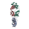
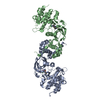
- Structure visualization
| Structure viewer | Molecule: MolmilJmol/JSmol |
|---|
- Downloads & links
-Download
| PDBx/mmCIF format | 1kvk.cif.gz | 85.2 KB | Display | PDBx/mmCIF format |
|---|---|---|---|---|
| PDB format | pdb1kvk.ent.gz | 63.9 KB | Display | PDB format |
| PDBx/mmJSON format | 1kvk.json.gz | Tree view | PDBx/mmJSON format | |
| Others |  Other downloads Other downloads |
-Validation report
| Arichive directory | https://data.pdbj.org/pub/pdb/validation_reports/kv/1kvkftp://data.pdbj.org/pub/pdb/validation_reports/kv/1kvk | HTTPS FTP |
|---|
-Related structure data
| Similar structure data |
|---|


-Links
 PDBj
PDBj- Assembly
| Deposited unit | 
| ||||||||
|---|---|---|---|---|---|---|---|---|---|
| 1 | 
| ||||||||
| Unit cell |
|
-Components
| #1: Protein | Mass: 42032.633 Da / Num. of mol.: 1 Source method: isolated from a genetically manipulated source Source: (gene. exp.)  |
|---|---|
| #2: Chemical | ChemComp-MG /   Mass: 24.305 Da / Num. of mol.: 1 / Source method: obtained synthetically / Formula: Mg Mass: 24.305 Da / Num. of mol.: 1 / Source method: obtained synthetically / Formula: Mg |
| #3: Chemical | ChemComp-ATP /   Mass: 507.181 Da / Num. of mol.: 1 / Source method: obtained synthetically / Formula: C10H16N5O13P3 / Comment: ATP, energy-carrying molecule*YM Mass: 507.181 Da / Num. of mol.: 1 / Source method: obtained synthetically / Formula: C10H16N5O13P3 / Comment: ATP, energy-carrying molecule*YM |
| #4: Water | ChemComp-HOH /  Mass: 18.015 Da / Num. of mol.: 63 / Source method: isolated from a natural source / Formula: H2O Mass: 18.015 Da / Num. of mol.: 63 / Source method: isolated from a natural source / Formula: H2O |
-Experimental details
-Experiment
| Experiment | Method: X-RAY DIFFRACTION / Number of used crystals: 1 |
|---|
- Sample preparation
| Crystal | Density Matthews: 2.38 Å3/Da / Density % sol: 48.25 % | ||||||||||||||||||||||||||||||||||||||||||
|---|---|---|---|---|---|---|---|---|---|---|---|---|---|---|---|---|---|---|---|---|---|---|---|---|---|---|---|---|---|---|---|---|---|---|---|---|---|---|---|---|---|---|---|
| Crystal grow | Temperature: 277 K / Method: vapor diffusion, sitting drop / pH: 7.5 Details: Hepes, MgCl2, ATP, PEG-5KMME, pH 7.5, VAPOR DIFFUSION, SITTING DROP, temperature 277K | ||||||||||||||||||||||||||||||||||||||||||
| Crystal grow | *PLUS Temperature: 4 ℃ | ||||||||||||||||||||||||||||||||||||||||||
| Components of the solutions | *PLUS
|
-Data collection
| Diffraction | Mean temperature: 277 K |
|---|---|
| Diffraction source | Source: ROTATING ANODE / Type: RIGAKU RU200 / Wavelength: 1.54 Å |
| Radiation | Protocol: SINGLE WAVELENGTH / Monochromatic (M) / Laue (L): M / Scattering type: x-ray |
| Radiation wavelength | Wavelength: 1.54 Å / Relative weight: 1 |
| Reflection | Resolution: 2.39→65.55 Å / Num. all: 14200 / Num. obs: 14200 / % possible obs: 86.9 % / Observed criterion σ(F): 0 / Observed criterion σ(I): 0 / Limit h max: 32 / Limit h min: 0 / Limit k max: 49 / Limit k min: 0 / Limit l max: 16 / Limit l min: 0 / Observed criterion F max: 282198.29 / Observed criterion F min: 0.55 |
| Reflection shell | Resolution: 2.4→2.49 Å / % possible all: 76.9 |
| Reflection | *PLUS Highest resolution: 2.4 Å / Num. obs: 14000 / Num. measured all: 30833 / Rmerge(I) obs: 0.054 |
| Reflection shell | *PLUS % possible obs: 77.8 % / Num. unique obs: 1011 / Num. measured obs: 2653 / Rmerge(I) obs: 0.305 |
- Processing
| Software |
| ||||||||||||||||||||||||||||||||||||||||||||||||||||||||||||||||||||||||||||||||||||||||||||||||||||||||||||||
|---|---|---|---|---|---|---|---|---|---|---|---|---|---|---|---|---|---|---|---|---|---|---|---|---|---|---|---|---|---|---|---|---|---|---|---|---|---|---|---|---|---|---|---|---|---|---|---|---|---|---|---|---|---|---|---|---|---|---|---|---|---|---|---|---|---|---|---|---|---|---|---|---|---|---|---|---|---|---|---|---|---|---|---|---|---|---|---|---|---|---|---|---|---|---|---|---|---|---|---|---|---|---|---|---|---|---|---|---|---|---|---|
| Refinement | Method to determine structure: MAD / Resolution: 2.4→65.5 Å / Rfactor Rfree error: 0.008 / Occupancy max: 1 / Occupancy min: 1 / Isotropic thermal model: Isotropic / Cross valid method: THROUGHOUT / σ(F): 0 / σ(I): 0
| ||||||||||||||||||||||||||||||||||||||||||||||||||||||||||||||||||||||||||||||||||||||||||||||||||||||||||||||
| Solvent computation | Solvent model: CNS bulk solvent model used / Bsol: 48.1123 Å2 / ksol: 0.301427 e/Å3 | ||||||||||||||||||||||||||||||||||||||||||||||||||||||||||||||||||||||||||||||||||||||||||||||||||||||||||||||
| Displacement parameters | Biso max: 88.5 Å2 / Biso mean: 43.09 Å2 / Biso min: 9.35 Å2 | ||||||||||||||||||||||||||||||||||||||||||||||||||||||||||||||||||||||||||||||||||||||||||||||||||||||||||||||
| Refine Biso |
| ||||||||||||||||||||||||||||||||||||||||||||||||||||||||||||||||||||||||||||||||||||||||||||||||||||||||||||||
| Refine analyze |
| ||||||||||||||||||||||||||||||||||||||||||||||||||||||||||||||||||||||||||||||||||||||||||||||||||||||||||||||
| Refinement step | Cycle: LAST / Resolution: 2.4→65.5 Å
| ||||||||||||||||||||||||||||||||||||||||||||||||||||||||||||||||||||||||||||||||||||||||||||||||||||||||||||||
| Refine LS restraints |
| ||||||||||||||||||||||||||||||||||||||||||||||||||||||||||||||||||||||||||||||||||||||||||||||||||||||||||||||
| LS refinement shell | Refine-ID: X-RAY DIFFRACTION
| ||||||||||||||||||||||||||||||||||||||||||||||||||||||||||||||||||||||||||||||||||||||||||||||||||||||||||||||
| Xplor file |
| ||||||||||||||||||||||||||||||||||||||||||||||||||||||||||||||||||||||||||||||||||||||||||||||||||||||||||||||
| Software | *PLUS Name: CNS / Classification: refinement | ||||||||||||||||||||||||||||||||||||||||||||||||||||||||||||||||||||||||||||||||||||||||||||||||||||||||||||||
| Refinement | *PLUS Highest resolution: 2.4 Å / Lowest resolution: 60 Å / Num. reflection obs: 14000 / % reflection Rfree: 8 % / Rfactor obs: 0.217 / Rfactor Rfree: 0.266 / Rfactor Rwork: 0.217 | ||||||||||||||||||||||||||||||||||||||||||||||||||||||||||||||||||||||||||||||||||||||||||||||||||||||||||||||
| Solvent computation | *PLUS | ||||||||||||||||||||||||||||||||||||||||||||||||||||||||||||||||||||||||||||||||||||||||||||||||||||||||||||||
| Displacement parameters | *PLUS | ||||||||||||||||||||||||||||||||||||||||||||||||||||||||||||||||||||||||||||||||||||||||||||||||||||||||||||||
| Refine LS restraints | *PLUS
| ||||||||||||||||||||||||||||||||||||||||||||||||||||||||||||||||||||||||||||||||||||||||||||||||||||||||||||||
| LS refinement shell | *PLUS Lowest resolution: 2.47 Å / Rfactor Rfree: 0.34 / Rfactor Rwork: 0.331 / Num. reflection Rwork: 1011 / Rfactor obs: 0.33 |
