 Movie
Movie Controller

[English] 日本語
 Yorodumi
- PDB-1kv0: Cis/trans Isomerization of Non-prolyl Peptide Bond Observed in Cr... -
+ Open data

- Basic information
| Entry | Database: PDB / ID: 1kv0 | ||||||
|---|---|---|---|---|---|---|---|
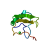
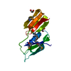
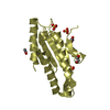
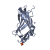
| Title | Cis/trans Isomerization of Non-prolyl Peptide Bond Observed in Crystal Structure of an Scorpion Toxin | ||||||
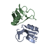
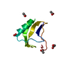
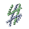
 Components Components | Alpha-like toxin BmK-M7 | ||||||
 Keywords Keywords | TOXIN / Non-prolyl cis peptide bond / Cis/trans isomerization / Dimeric structure / Scorpion toxin | ||||||
| Function / homology |  Function and homology information Function and homology informationsodium channel inhibitor activity / defense response / toxin activity / extracellular region Similarity search - Function | ||||||
| Biological species |  Mesobuthus martensii (Chinese scorpion) Mesobuthus martensii (Chinese scorpion) | ||||||
| Method |  X-RAY DIFFRACTION / SYNCHROTRON / MOLECULAR REPLACEMENT / Resolution: 1.4 Å X-RAY DIFFRACTION / SYNCHROTRON / MOLECULAR REPLACEMENT / Resolution: 1.4 Å | ||||||
 Authors Authors | Guan, R.J. / He, X.L. / Wang, M. / Xiang, Y. / Wang, D.C. | ||||||
 Citation Citation | Journal: J.Mol.Biol. / Year: 2004 Title: Structural mechanism governing cis and trans isomeric states and an intramolecular switch for cis/trans isomerization of a non-proline peptide bond observed in crystal structures of scorpion toxins. Authors: Guan, R.J. / Xiang, Y. / He, X.L. / Wang, C.G. / Wang, M. / Zhang, Y. / Sundberg, E.J. / Wang, D.C. | ||||||
| History |
|
- Structure visualization
| Structure viewer | Molecule: MolmilJmol/JSmol |
|---|
- Downloads & links
-Download
| PDBx/mmCIF format | 1kv0.cif.gz | 67.3 KB | Display | PDBx/mmCIF format |
|---|---|---|---|---|
| PDB format | pdb1kv0.ent.gz | 51 KB | Display | PDB format |
| PDBx/mmJSON format | 1kv0.json.gz | Tree view | PDBx/mmJSON format | |
| Others |  Other downloads Other downloads |
-Validation report
| Arichive directory | https://data.pdbj.org/pub/pdb/validation_reports/kv/1kv0ftp://data.pdbj.org/pub/pdb/validation_reports/kv/1kv0 | HTTPS FTP |
|---|
-Related structure data
| Related structure data | 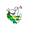 1t7aC 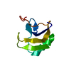 1t7bC  1t7eC 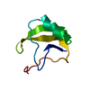 1sn1S S: Starting model for refinement C: citing same article ( |
|---|---|
| Similar structure data |


-Links
 PDBj
PDBj

- Assembly
| Deposited unit | 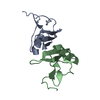
| |||||||||||||||
|---|---|---|---|---|---|---|---|---|---|---|---|---|---|---|---|---|
| 1 |
| |||||||||||||||
| Unit cell |
| |||||||||||||||
| Components on special symmetry positions |
|
-Components
| #1: Protein | Mass: 7252.350 Da / Num. of mol.: 2 / Source method: isolated from a natural source / Source: (natural) Mesobuthus martensii (Chinese scorpion) / Tissue: tail venom gland / References: UniProt: P59854#2: Water | ChemComp-HOH / |  Mass: 18.015 Da / Num. of mol.: 118 / Source method: isolated from a natural source / Formula: H2O Mass: 18.015 Da / Num. of mol.: 118 / Source method: isolated from a natural source / Formula: H2OHas protein modification | Y | |
|---|
-Experimental details
-Experiment
| Experiment | Method: X-RAY DIFFRACTION / Number of used crystals: 1 |
|---|
- Sample preparation
| Crystal | Density Matthews: 1.89 Å3/Da / Density % sol: 34.81 % | ||||||||||||||||||||||||||||||
|---|---|---|---|---|---|---|---|---|---|---|---|---|---|---|---|---|---|---|---|---|---|---|---|---|---|---|---|---|---|---|---|
| Crystal grow | Temperature: 293 K / Method: vapor diffusion, hanging drop / pH: 8.5 Details: ammonium sulfate, Tris-HCl, Ethanol, pH 8.5, VAPOR DIFFUSION, HANGING DROP, temperature 293K | ||||||||||||||||||||||||||||||
| Crystal grow | *PLUS Method: vapor diffusion, hanging drop | ||||||||||||||||||||||||||||||
| Components of the solutions | *PLUS
|
-Data collection
| Diffraction | Mean temperature: 293 K |
|---|---|
| Diffraction source | Source: SYNCHROTRON / Site: Photon Factory  / Beamline: BL-18B / Wavelength: 1 Å / Beamline: BL-18B / Wavelength: 1 Å |
| Detector | Type: ADSC QUANTUM 4 / Detector: CCD / Date: Dec 24, 2000 |
| Radiation | Protocol: SINGLE WAVELENGTH / Monochromatic (M) / Laue (L): M / Scattering type: x-ray |
| Radiation wavelength | Wavelength: 1 Å / Relative weight: 1 |
| Reflection | Resolution: 1.4→29.3 Å / Num. obs: 19260 / % possible obs: 85.8 % / Observed criterion σ(F): 0 / Rmerge(I) obs: 0.04 |
| Reflection shell | Resolution: 1.4→1.48 Å / Rmerge(I) obs: 0.201 / Mean I/σ(I) obs: 3.6 / % possible all: 74.6 |
| Reflection | *PLUS Highest resolution: 1.4 Å / Num. measured all: 57131 / Rmerge(I) obs: 0.04 |
| Reflection shell | *PLUS % possible obs: 74.6 % |
- Processing
| Software |
| |||||||||||||||||||||||||||||||||||||||||||||||||||||||||||||||||||||||||||||||||||||||||||||||||||||||||||||||||||||||||||||
|---|---|---|---|---|---|---|---|---|---|---|---|---|---|---|---|---|---|---|---|---|---|---|---|---|---|---|---|---|---|---|---|---|---|---|---|---|---|---|---|---|---|---|---|---|---|---|---|---|---|---|---|---|---|---|---|---|---|---|---|---|---|---|---|---|---|---|---|---|---|---|---|---|---|---|---|---|---|---|---|---|---|---|---|---|---|---|---|---|---|---|---|---|---|---|---|---|---|---|---|---|---|---|---|---|---|---|---|---|---|---|---|---|---|---|---|---|---|---|---|---|---|---|---|---|---|---|
| Refinement | Method to determine structure: MOLECULAR REPLACEMENT Starting model: PDB entry 1SN1 Resolution: 1.4→29.49 Å / Cor.coef. Fo:Fc: 0.969 / Cor.coef. Fo:Fc free: 0.964 / SU B: 0.795 / SU ML: 0.032 / Cross valid method: THROUGHOUT / σ(F): 0 / ESU R: 0.084 / ESU R Free: 0.062 / Stereochemistry target values: MAXIMUM LIKELIHOOD / Details: HYDROGENS HAVE BEEN ADDED IN THE RIDING POSITIONS
| |||||||||||||||||||||||||||||||||||||||||||||||||||||||||||||||||||||||||||||||||||||||||||||||||||||||||||||||||||||||||||||
| Solvent computation | Ion probe radii: 0.8 Å / Shrinkage radii: 0.8 Å / VDW probe radii: 1.4 Å | |||||||||||||||||||||||||||||||||||||||||||||||||||||||||||||||||||||||||||||||||||||||||||||||||||||||||||||||||||||||||||||
| Displacement parameters | Biso mean: 15.774 Å2
| |||||||||||||||||||||||||||||||||||||||||||||||||||||||||||||||||||||||||||||||||||||||||||||||||||||||||||||||||||||||||||||
| Refinement step | Cycle: LAST / Resolution: 1.4→29.49 Å
| |||||||||||||||||||||||||||||||||||||||||||||||||||||||||||||||||||||||||||||||||||||||||||||||||||||||||||||||||||||||||||||
| Refine LS restraints |
| |||||||||||||||||||||||||||||||||||||||||||||||||||||||||||||||||||||||||||||||||||||||||||||||||||||||||||||||||||||||||||||
| LS refinement shell | Resolution: 1.4→1.436 Å / Total num. of bins used: 20
| |||||||||||||||||||||||||||||||||||||||||||||||||||||||||||||||||||||||||||||||||||||||||||||||||||||||||||||||||||||||||||||
| Refinement | *PLUS Lowest resolution: 29.3 Å / % reflection Rfree: 10 % / Rfactor Rfree: 0.164 / Rfactor Rwork: 0.142 | |||||||||||||||||||||||||||||||||||||||||||||||||||||||||||||||||||||||||||||||||||||||||||||||||||||||||||||||||||||||||||||
| Solvent computation | *PLUS | |||||||||||||||||||||||||||||||||||||||||||||||||||||||||||||||||||||||||||||||||||||||||||||||||||||||||||||||||||||||||||||
| Displacement parameters | *PLUS | |||||||||||||||||||||||||||||||||||||||||||||||||||||||||||||||||||||||||||||||||||||||||||||||||||||||||||||||||||||||||||||
| Refine LS restraints | *PLUS
|
