 Movie
Movie Controller


+ Open data

- Basic information
| Entry | Database: PDB / ID: 1kuh | ||||||
|---|---|---|---|---|---|---|---|
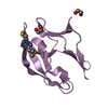
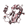
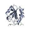
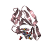
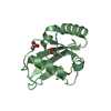
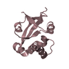
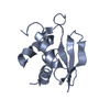
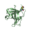
| Title | ZINC PROTEASE FROM STREPTOMYCES CAESPITOSUS | ||||||
 Components Components | ZINC PROTEASE | ||||||
 Keywords Keywords | HYDROLASE / METALLOPROTEINASE | ||||||
| Function / homology |  Function and homology information Function and homology informationsnapalysin / metalloendopeptidase activity / proteolysis / extracellular region / zinc ion binding Similarity search - Function | ||||||
| Biological species |  Streptomyces caespitosus (bacteria) Streptomyces caespitosus (bacteria) | ||||||
| Method |  X-RAY DIFFRACTION / Resolution: 1.6 Å X-RAY DIFFRACTION / Resolution: 1.6 Å | ||||||
 Authors Authors | Kurisu, G. / Kinoshita, T. / Sugimoto, A. / Nagara, A. / Kai, Y. / Kasai, N. / Harada, S. | ||||||
 Citation Citation | Journal: J.Biochem.(Tokyo) / Year: 1997 Title: Structure of the zinc endoprotease from Streptomyces caespitosus. Authors: Kurisu, G. / Kinoshita, T. / Sugimoto, A. / Nagara, A. / Kai, Y. / Kasai, N. / Harada, S. #1: Journal: Eur.J.Biochem. / Year: 1995Title: Complete Amino Acid Sequence of a Zinc Metalloendoprotease from Streptomyces Caespitosus Authors: Harada, S. / Kinoshita, T. / Kasai, N. / Tsunasawa, S. / Sakiyama, F. #2: Journal: J.Biochem.(Tokyo) / Year: 1991Title: Crystallization and Main-Chain Structure of Neutral Protease from Streptomyces Caespitosus Authors: Harada, S. / Kitadokoro, K. / Kinoshita, T. / Kai, Y. / Kasai, N. | ||||||
| History |
|
- Structure visualization
| Structure viewer | Molecule: MolmilJmol/JSmol |
|---|
- Downloads & links
-Download
| PDBx/mmCIF format | 1kuh.cif.gz | 39.2 KB | Display | PDBx/mmCIF format |
|---|---|---|---|---|
| PDB format | pdb1kuh.ent.gz | 26.7 KB | Display | PDB format |
| PDBx/mmJSON format | 1kuh.json.gz | Tree view | PDBx/mmJSON format | |
| Others |  Other downloads Other downloads |
-Validation report
| Arichive directory | https://data.pdbj.org/pub/pdb/validation_reports/ku/1kuhftp://data.pdbj.org/pub/pdb/validation_reports/ku/1kuh | HTTPS FTP |
|---|
-Related structure data
| Similar structure data |
|---|


-Links
 PDBj
PDBj


- Assembly
| Deposited unit | 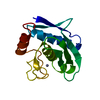
| ||||||||
|---|---|---|---|---|---|---|---|---|---|
| 1 |
| ||||||||
| Unit cell |
|
-Components
| #1: Protein | Mass: 14387.327 Da / Num. of mol.: 1 / Source method: isolated from a natural source / Source: (natural) Streptomyces caespitosus (bacteria) / References: UniProt: P56406 |
|---|---|
| #2: Chemical | ChemComp-ZN /   Mass: 65.409 Da / Num. of mol.: 1 / Source method: obtained synthetically / Formula: Zn Mass: 65.409 Da / Num. of mol.: 1 / Source method: obtained synthetically / Formula: Zn |
| #3: Chemical | ChemComp-CA /   Mass: 40.078 Da / Num. of mol.: 1 / Source method: obtained synthetically / Formula: Ca Mass: 40.078 Da / Num. of mol.: 1 / Source method: obtained synthetically / Formula: Ca |
| #4: Water | ChemComp-HOH /  Mass: 18.015 Da / Num. of mol.: 93 / Source method: isolated from a natural source / Formula: H2O Mass: 18.015 Da / Num. of mol.: 93 / Source method: isolated from a natural source / Formula: H2O |
| Has protein modification | Y |
-Experimental details
-Experiment
| Experiment | Method: X-RAY DIFFRACTION |
|---|
- Sample preparation
| Crystal | Density Matthews: 1.99 Å3/Da / Density % sol: 35 % | ||||||||||||||||||||
|---|---|---|---|---|---|---|---|---|---|---|---|---|---|---|---|---|---|---|---|---|---|
| Crystal grow | *PLUS Temperature: 4 ℃ / Method: batch method | ||||||||||||||||||||
| Components of the solutions | *PLUS
|
-Data collection
| Diffraction source | Wavelength: 1.5418 |
|---|---|
| Detector | Type: RIGAKU / Detector: IMAGE PLATE |
| Radiation | Monochromatic (M) / Laue (L): M / Scattering type: x-ray |
| Radiation wavelength | Wavelength: 1.5418 Å / Relative weight: 1 |
| Reflection | Num. obs: 19872 / Observed criterion σ(I): 2 |
- Processing
| Software | Name: PROLSQ / Classification: refinement | ||||||||||||||||||||||||||||||||||||||||||||||||||||||||||||||||||||||||||||||||||||
|---|---|---|---|---|---|---|---|---|---|---|---|---|---|---|---|---|---|---|---|---|---|---|---|---|---|---|---|---|---|---|---|---|---|---|---|---|---|---|---|---|---|---|---|---|---|---|---|---|---|---|---|---|---|---|---|---|---|---|---|---|---|---|---|---|---|---|---|---|---|---|---|---|---|---|---|---|---|---|---|---|---|---|---|---|---|
| Refinement | Resolution: 1.6→10 Å / σ(F): 2 /
| ||||||||||||||||||||||||||||||||||||||||||||||||||||||||||||||||||||||||||||||||||||
| Displacement parameters | Biso mean: 10.45 Å2 | ||||||||||||||||||||||||||||||||||||||||||||||||||||||||||||||||||||||||||||||||||||
| Refinement step | Cycle: LAST / Resolution: 1.6→10 Å
| ||||||||||||||||||||||||||||||||||||||||||||||||||||||||||||||||||||||||||||||||||||
| Refine LS restraints |
| ||||||||||||||||||||||||||||||||||||||||||||||||||||||||||||||||||||||||||||||||||||
| Refinement | *PLUS Num. reflection obs: 12926 / Rfactor obs: 0.166 | ||||||||||||||||||||||||||||||||||||||||||||||||||||||||||||||||||||||||||||||||||||
| Solvent computation | *PLUS | ||||||||||||||||||||||||||||||||||||||||||||||||||||||||||||||||||||||||||||||||||||
| Displacement parameters | *PLUS | ||||||||||||||||||||||||||||||||||||||||||||||||||||||||||||||||||||||||||||||||||||
| Refine LS restraints | *PLUS
|
