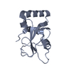
 Movie
Movie Controller


+ Open data

- Basic information
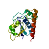
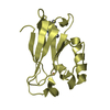
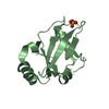
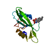
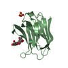
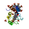
| Entry | Database: PDB / ID: 6hoi | |||||||||
|---|---|---|---|---|---|---|---|---|---|---|
| Title | Structure of Beclin1 LIR motif bound to GABARAPL1 | |||||||||
 Components Components |
| |||||||||
 Keywords Keywords | SIGNALING PROTEIN / Autophagy / ATG8 / LIR | |||||||||
| Function / homology |  Function and homology information Function and homology informationcellular response to aluminum ion / cargo adaptor activity / positive regulation of stress granule assembly / phosphatidylinositol 3-kinase complex, class III / cellular response to oxygen-glucose deprivation / phosphatidylinositol 3-kinase complex, class III, type II / phosphatidylinositol 3-kinase complex, class III, type I / glycophagy / response to mitochondrial depolarisation / positive regulation of attachment of mitotic spindle microtubules to kinetochore ...cellular response to aluminum ion / cargo adaptor activity / positive regulation of stress granule assembly / phosphatidylinositol 3-kinase complex, class III / cellular response to oxygen-glucose deprivation / phosphatidylinositol 3-kinase complex, class III, type II / phosphatidylinositol 3-kinase complex, class III, type I / glycophagy / response to mitochondrial depolarisation / positive regulation of attachment of mitotic spindle microtubules to kinetochore / engulfment of apoptotic cell / negative regulation of lysosome organization / positive regulation of autophagosome assembly / cytoplasmic side of mitochondrial outer membrane / early endosome to late endosome transport / negative regulation of autophagosome assembly / receptor catabolic process / SMAD protein signal transduction / late endosome to vacuole transport / protein targeting to lysosome / GABA receptor binding / phosphatidylethanolamine binding / Tat protein binding / phagophore assembly site / Translation of Replicase and Assembly of the Replication Transcription Complex / cellular response to nitrogen starvation / phosphatidylinositol-3-phosphate biosynthetic process / negative regulation of programmed cell death / mitotic metaphase chromosome alignment / response to vitamin E / lysosome organization / Macroautophagy / response to iron(II) ion / positive regulation of cardiac muscle hypertrophy / RSV-host interactions / p38MAPK cascade / cytoplasmic pattern recognition receptor signaling pathway / autophagosome membrane / amyloid-beta metabolic process / protein secretion / regulation of macroautophagy / autophagosome maturation / neuron development / cellular defense response / autophagosome assembly / phosphatidylinositol 3-kinase binding / beta-tubulin binding / cellular response to glucose starvation / mitophagy / JNK cascade / phagocytic vesicle / positive regulation of intrinsic apoptotic signaling pathway / positive regulation of autophagy / autophagosome / cellular response to epidermal growth factor stimulus / cytoplasmic vesicle membrane / cellular response to copper ion / cellular response to amino acid starvation / regulation of autophagy / regulation of cytokinesis / Antigen Presentation: Folding, assembly and peptide loading of class I MHC / macroautophagy / trans-Golgi network / circadian rhythm / phospholipid binding / bone development / response to lead ion / autophagy / ISG15 antiviral mechanism / cellular response to hydrogen peroxide / protein-containing complex assembly / GTPase binding / Translation of Replicase and Assembly of the Replication Transcription Complex / defense response to virus / molecular adaptor activity / microtubule / protein-macromolecule adaptor activity / response to hypoxia / positive regulation of phosphatidylinositol 3-kinase/protein kinase B signal transduction / endosome / endosome membrane / Ub-specific processing proteases / cilium / nuclear body / ciliary basal body / response to xenobiotic stimulus / negative regulation of cell population proliferation / cell division / apoptotic process / ubiquitin protein ligase binding / dendrite / endoplasmic reticulum membrane / protein kinase binding / negative regulation of apoptotic process / SARS-CoV-2 activates/modulates innate and adaptive immune responses / Golgi apparatus / endoplasmic reticulum / identical protein binding / plasma membrane / cytoplasm Similarity search - Function | |||||||||
| Biological species |  Homo sapiens (human) Homo sapiens (human) | |||||||||
| Method |  X-RAY DIFFRACTION / SYNCHROTRON / MOLECULAR REPLACEMENT / Resolution: 1.14 Å X-RAY DIFFRACTION / SYNCHROTRON / MOLECULAR REPLACEMENT / Resolution: 1.14 Å | |||||||||
 Authors Authors | Mouilleron, S. / Birgisdottir, A.B. / Bhujbal, Z. / Wirth, M. / Sjottem, E. / Evjen, G. / Zhang, W. / Lee, R. / O'Reilly, N. / Tooze, S. ...Mouilleron, S. / Birgisdottir, A.B. / Bhujbal, Z. / Wirth, M. / Sjottem, E. / Evjen, G. / Zhang, W. / Lee, R. / O'Reilly, N. / Tooze, S. / Lamark, T. / Johansen, T. | |||||||||
| Funding support |  Norway, 2items Norway, 2items
| |||||||||
 Citation Citation | Journal: Autophagy / Year: 2019 Title: Members of the autophagy class III phosphatidylinositol 3-kinase complex I interact with GABARAP and GABARAPL1 via LIR motifs. Authors: Birgisdottir, A.B. / Mouilleron, S. / Bhujabal, Z. / Wirth, M. / Sjottem, E. / Evjen, G. / Zhang, W. / Lee, R. / O'Reilly, N. / Tooze, S.A. / Lamark, T. / Johansen, T. | |||||||||
| History |
|
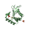
- Structure visualization
| Structure viewer | Molecule: MolmilJmol/JSmol |
|---|
- Downloads & links
-Download
| PDBx/mmCIF format | 6hoi.cif.gz | 128 KB | Display | PDBx/mmCIF format |
|---|---|---|---|---|
| PDB format | pdb6hoi.ent.gz | 99.9 KB | Display | PDB format |
| PDBx/mmJSON format | 6hoi.json.gz | Tree view | PDBx/mmJSON format | |
| Others |  Other downloads Other downloads |
-Validation report
| Arichive directory | https://data.pdbj.org/pub/pdb/validation_reports/ho/6hoiftp://data.pdbj.org/pub/pdb/validation_reports/ho/6hoi | HTTPS FTP |
|---|
-Related structure data
| Related structure data | 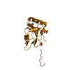 6hogC 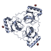 6hohC 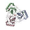 6hojC 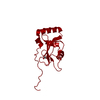 6hokC 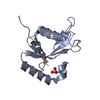 6holC 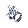 2r2qS S: Starting model for refinement C: citing same article ( |
|---|---|
| Similar structure data |

-Links
 PDBj
PDBj


- Assembly
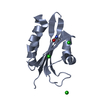
| Deposited unit | 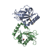
| ||||||||
|---|---|---|---|---|---|---|---|---|---|
| 1 | 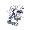
| ||||||||
| 2 | 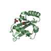
| ||||||||
| Unit cell |
|
-Components
| #1: Protein | Mass: 14598.667 Da / Num. of mol.: 2 Source method: isolated from a genetically manipulated source Source: (gene. exp.) Homo sapiens (human) / Gene: GABARAPL1, GEC1 / Production host:  #2: Protein/peptide | Mass: 1038.110 Da / Num. of mol.: 2 / Source method: obtained synthetically / Source: (synth.) Homo sapiens (human) / References: UniProt: Q14457#3: Chemical | ChemComp-EDO / |   Mass: 62.068 Da / Num. of mol.: 1 / Source method: obtained synthetically / Formula: C2H6O2 Mass: 62.068 Da / Num. of mol.: 1 / Source method: obtained synthetically / Formula: C2H6O2#4: Chemical |   Mass: 59.044 Da / Num. of mol.: 3 / Source method: obtained synthetically / Formula: C2H3O2 Mass: 59.044 Da / Num. of mol.: 3 / Source method: obtained synthetically / Formula: C2H3O2#5: Water | ChemComp-HOH / |  Mass: 18.015 Da / Num. of mol.: 183 / Source method: isolated from a natural source / Formula: H2O Mass: 18.015 Da / Num. of mol.: 183 / Source method: isolated from a natural source / Formula: H2O |
|---|
-Experimental details
-Experiment
| Experiment | Method: X-RAY DIFFRACTION / Number of used crystals: 1 |
|---|
- Sample preparation
| Crystal | Density Matthews: 1.94 Å3/Da / Density % sol: 36.65 % |
|---|---|
| Crystal grow | Temperature: 293 K / Method: vapor diffusion, sitting drop Details: 0.1M sodium acetate, 0.1M Hepes pH 7.5, 12% PEG 4000 |
-Data collection
| Diffraction | Mean temperature: 100 K |
|---|---|
| Diffraction source | Source: SYNCHROTRON / Site: Diamond  / Beamline: I02 / Wavelength: 0.97 Å / Beamline: I02 / Wavelength: 0.97 Å |
| Detector | Type: DECTRIS PILATUS3 6M / Detector: PIXEL / Date: Jul 17, 2015 |
| Radiation | Protocol: SINGLE WAVELENGTH / Monochromatic (M) / Laue (L): M / Scattering type: x-ray |
| Radiation wavelength | Wavelength: 0.97 Å / Relative weight: 1 |
| Reflection | Resolution: 1.14→35.161 Å / Num. obs: 88324 / % possible obs: 99.34 % / Redundancy: 4.1 % / Biso Wilson estimate: 14 Å2 / CC1/2: 0.99 / Rmerge(I) obs: 0.03 / Rpim(I) all: 0.01 / Rrim(I) all: 0.03 / Net I/σ(I): 18.2 |
| Reflection shell | Resolution: 1.14→1.18 Å / Redundancy: 2.8 % / Rmerge(I) obs: 0.64 / Mean I/σ(I) obs: 1.7 / Num. unique obs: 8545 / CC1/2: 0.58 / Rpim(I) all: 0.44 / Rrim(I) all: 0.78 / % possible all: 97.51 |
- Processing
| Software |
| |||||||||||||||||||||||||||||||||||||||||||||||||||||||||||||||||||||||||||||||||||||||||||||||||||||||||||||||||||||||||||||||||||||||||||||||||||||||||||||||||||||||||||||||||||||||||||||||||||||||||||||||||||||||||
|---|---|---|---|---|---|---|---|---|---|---|---|---|---|---|---|---|---|---|---|---|---|---|---|---|---|---|---|---|---|---|---|---|---|---|---|---|---|---|---|---|---|---|---|---|---|---|---|---|---|---|---|---|---|---|---|---|---|---|---|---|---|---|---|---|---|---|---|---|---|---|---|---|---|---|---|---|---|---|---|---|---|---|---|---|---|---|---|---|---|---|---|---|---|---|---|---|---|---|---|---|---|---|---|---|---|---|---|---|---|---|---|---|---|---|---|---|---|---|---|---|---|---|---|---|---|---|---|---|---|---|---|---|---|---|---|---|---|---|---|---|---|---|---|---|---|---|---|---|---|---|---|---|---|---|---|---|---|---|---|---|---|---|---|---|---|---|---|---|---|---|---|---|---|---|---|---|---|---|---|---|---|---|---|---|---|---|---|---|---|---|---|---|---|---|---|---|---|---|---|---|---|---|---|---|---|---|---|---|---|---|---|---|---|---|---|---|---|---|
| Refinement | Method to determine structure: MOLECULAR REPLACEMENT Starting model: 2R2Q Resolution: 1.14→35.161 Å / SU ML: 0.11 / Cross valid method: FREE R-VALUE / σ(F): 1.34 / Phase error: 15.32
| |||||||||||||||||||||||||||||||||||||||||||||||||||||||||||||||||||||||||||||||||||||||||||||||||||||||||||||||||||||||||||||||||||||||||||||||||||||||||||||||||||||||||||||||||||||||||||||||||||||||||||||||||||||||||
| Solvent computation | Shrinkage radii: 0.9 Å / VDW probe radii: 1.11 Å | |||||||||||||||||||||||||||||||||||||||||||||||||||||||||||||||||||||||||||||||||||||||||||||||||||||||||||||||||||||||||||||||||||||||||||||||||||||||||||||||||||||||||||||||||||||||||||||||||||||||||||||||||||||||||
| Refinement step | Cycle: LAST / Resolution: 1.14→35.161 Å
| |||||||||||||||||||||||||||||||||||||||||||||||||||||||||||||||||||||||||||||||||||||||||||||||||||||||||||||||||||||||||||||||||||||||||||||||||||||||||||||||||||||||||||||||||||||||||||||||||||||||||||||||||||||||||
| Refine LS restraints |
| |||||||||||||||||||||||||||||||||||||||||||||||||||||||||||||||||||||||||||||||||||||||||||||||||||||||||||||||||||||||||||||||||||||||||||||||||||||||||||||||||||||||||||||||||||||||||||||||||||||||||||||||||||||||||
| LS refinement shell |
|
