 Movie
Movie Controller


+ Open data

- Basic information
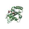
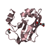
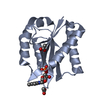
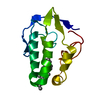
| Entry | Database: PDB / ID: 1c7k | ||||||
|---|---|---|---|---|---|---|---|
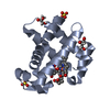
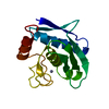
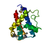
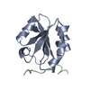
| Title | CRYSTAL STRUCTURE OF THE ZINC PROTEASE | ||||||
 Components Components | ZINC ENDOPROTEASE | ||||||
 Keywords Keywords | HYDROLASE / alpha and beta protein / METALLOPROTEINASE | ||||||
| Function / homology |  Function and homology information Function and homology informationsnapalysin / metalloendopeptidase activity / proteolysis / extracellular region / zinc ion binding Similarity search - Function | ||||||
| Biological species |  Streptomyces caespitosus (bacteria) Streptomyces caespitosus (bacteria) | ||||||
| Method |  X-RAY DIFFRACTION / AB INITIO / Resolution: 1 Å X-RAY DIFFRACTION / AB INITIO / Resolution: 1 Å | ||||||
 Authors Authors | Kurisu, G. / Harada, S. / Kai, Y. | ||||||
 Citation Citation | Journal: J.Inorg.Biochem. / Year: 2000 Title: Structure of the zinc-binding site in the crystal structure of a zinc endoprotease from Streptomyces caespitosus at 1 A resolution. Authors: Kurisu, G. / Kai, Y. / Harada, S. #1: Journal: J.BIOCHEM.(TOKYO) / Year: 1997Title: Structure of the Zinc Endoprotease from Streptomyces caespitosus Authors: Kurisu, G. / Kinoshita, T. / Sugimoto, A. / Nagara, A. / Kai, Y. / Kasai, N. / Harada, S. #2: Journal: Eur.J.Biochem. / Year: 1995Title: Complete Amino Acid Sequence of a Zinc Metalloendoprotease from Streptomyces caespitosus Authors: Harada, S. / Kinoshita, T. / Kasai, N. / Tsunasawa, S. / Sakiyama, F. #3: Journal: J.BIOCHEM.(TOKYO) / Year: 1991Title: Crystallization and Main-Chain Structure of Neutral Protease from Streptomyces caespitosus Authors: Harada, S. / Kitadokoro, K. / Kinoshita, T. / Kai, Y. / Kasai, N. | ||||||
| History |
|
- Structure visualization
| Structure viewer | Molecule: MolmilJmol/JSmol |
|---|
- Downloads & links
-Download
| PDBx/mmCIF format | 1c7k.cif.gz | 66.9 KB | Display | PDBx/mmCIF format |
|---|---|---|---|---|
| PDB format | pdb1c7k.ent.gz | 48.5 KB | Display | PDB format |
| PDBx/mmJSON format | 1c7k.json.gz | Tree view | PDBx/mmJSON format | |
| Others |  Other downloads Other downloads |
-Validation report
| Arichive directory | https://data.pdbj.org/pub/pdb/validation_reports/c7/1c7kftp://data.pdbj.org/pub/pdb/validation_reports/c7/1c7k | HTTPS FTP |
|---|
-Related structure data
| Similar structure data |
|---|


-Links
 PDBj
PDBj


- Assembly
| Deposited unit | 
| ||||||||
|---|---|---|---|---|---|---|---|---|---|
| 1 |
| ||||||||
| Unit cell |
|
-Components
| #1: Protein | Mass: 14387.327 Da / Num. of mol.: 1 / Source method: isolated from a natural source / Source: (natural) Streptomyces caespitosus (bacteria)References: UniProt: P56406, Hydrolases; Acting on peptide bonds (peptidases); Metalloendopeptidases |
|---|---|
| #2: Chemical | ChemComp-ZN /   Mass: 65.409 Da / Num. of mol.: 1 / Source method: obtained synthetically / Formula: Zn Mass: 65.409 Da / Num. of mol.: 1 / Source method: obtained synthetically / Formula: Zn |
| #3: Chemical | ChemComp-CA /   Mass: 40.078 Da / Num. of mol.: 1 / Source method: obtained synthetically / Formula: Ca Mass: 40.078 Da / Num. of mol.: 1 / Source method: obtained synthetically / Formula: Ca |
| #4: Water | ChemComp-HOH /  Mass: 18.015 Da / Num. of mol.: 116 / Source method: isolated from a natural source / Formula: H2O Mass: 18.015 Da / Num. of mol.: 116 / Source method: isolated from a natural source / Formula: H2O |
| Has protein modification | Y |
-Experimental details
-Experiment
| Experiment | Method: X-RAY DIFFRACTION / Number of used crystals: 1 |
|---|
- Sample preparation
| Crystal | Density Matthews: 1 Å3/Da / Density % sol: 37.48 % | ||||||||||||||||||||
|---|---|---|---|---|---|---|---|---|---|---|---|---|---|---|---|---|---|---|---|---|---|
| Crystal grow | Temperature: 277 K / Method: liquid diffusion / pH: 7 Details: acetone, pH 7.0, LIQUID DIFFUSION, temperature 277K | ||||||||||||||||||||
| Crystal | *PLUS Density % sol: 35 % | ||||||||||||||||||||
| Crystal grow | *PLUS Temperature: 4 ℃ / Method: batch method / Details: Harada, S., (1991) J.BIOCHEM.(TOKYO), 110, 46. | ||||||||||||||||||||
| Components of the solutions | *PLUS
|
-Data collection
| Diffraction | Mean temperature: 277 K |
|---|---|
| Diffraction source | Source: ROTATING ANODE / Type: RIGAKU RU300 / Wavelength: 0.7107 |
| Detector | Type: RIGAKU RAXIS / Detector: IMAGE PLATE / Date: Dec 18, 1997 |
| Radiation | Protocol: SINGLE WAVELENGTH / Monochromatic (M) / Laue (L): M / Scattering type: x-ray |
| Radiation wavelength | Wavelength: 0.7107 Å / Relative weight: 1 |
| Reflection | Resolution: 0.92→55 Å / Num. all: 59541 / Num. obs: 59541 / % possible obs: 81.9 % / Observed criterion σ(I): 0 / Rmerge(I) obs: 0.057 / Net I/σ(I): 14.64 |
| Reflection shell | Resolution: 0.92→0.97 Å / % possible all: 58.3 |
| Reflection | *PLUS Num. obs: 51419 / % possible obs: 82 % / Redundancy: 58.3 % / Num. measured all: 158614 |
- Processing
| Software |
| |||||||||||||||||||||||||||||||||
|---|---|---|---|---|---|---|---|---|---|---|---|---|---|---|---|---|---|---|---|---|---|---|---|---|---|---|---|---|---|---|---|---|---|---|
| Refinement | Method to determine structure: AB INITIO / Resolution: 1→55 Å / Num. parameters: 992 / Num. restraintsaints: 1187 / Cross valid method: FREE R / σ(F): 0 / Stereochemistry target values: Engh & Huber
| |||||||||||||||||||||||||||||||||
| Refine analyze | Occupancy sum hydrogen: 0 / Occupancy sum non hydrogen: 1133 | |||||||||||||||||||||||||||||||||
| Refinement step | Cycle: LAST / Resolution: 1→55 Å
| |||||||||||||||||||||||||||||||||
| Refine LS restraints |
| |||||||||||||||||||||||||||||||||
| Software | *PLUS Name: SHELXL-97 / Classification: refinement | |||||||||||||||||||||||||||||||||
| Refinement | *PLUS Num. reflection all: 51419 / Rfactor Rfree: 0.17 / Rfactor Rwork: 0.15 | |||||||||||||||||||||||||||||||||
| Solvent computation | *PLUS | |||||||||||||||||||||||||||||||||
| Displacement parameters | *PLUS | |||||||||||||||||||||||||||||||||
| Refine LS restraints | *PLUS
|
