 Movie
Movie Controller

[English] 日本語
 Yorodumi
- PDB-1kkr: CRYSTAL STRUCTURE OF CITROBACTER AMALONATICUS METHYLASPARTATE AMM... -
+ Open data

- Basic information
| Entry | Database: PDB / ID: 1kkr | ||||||
|---|---|---|---|---|---|---|---|
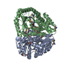
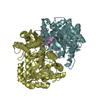
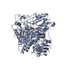
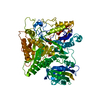
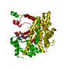
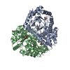
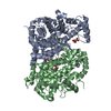
| Title | CRYSTAL STRUCTURE OF CITROBACTER AMALONATICUS METHYLASPARTATE AMMONIA LYASE CONTAINING (2S,3S)-3-METHYLASPARTIC ACID | ||||||
 Components Components | 3-METHYLASPARTATE AMMONIA-LYASE | ||||||
 Keywords Keywords | LYASE / Methylaspartate ammonia lyase / Enolase superfamily / TIM barrel / substrate complex | ||||||
| Function / homology |  Function and homology information Function and homology informationmethylaspartate ammonia-lyase / methylaspartate ammonia-lyase activity / : / metal ion binding Similarity search - Function | ||||||
| Biological species |  Citrobacter amalonaticus (bacteria) Citrobacter amalonaticus (bacteria) | ||||||
| Method |  X-RAY DIFFRACTION / Resolution: 2.1 Å X-RAY DIFFRACTION / Resolution: 2.1 Å | ||||||
 Authors Authors | Levy, C.W. / Buckley, P.A. / Sedelnikova, S. / Kato, K. / Asano, Y. / Rice, D.W. / Baker, P.J. | ||||||
 Citation Citation | Journal: Structure / Year: 2002 Title: Insights into enzyme evolution revealed by the structure of methylaspartate ammonia lyase. Authors: Levy, C.W. / Buckley, P.A. / Sedelnikova, S. / Kato, Y. / Asano, Y. / Rice, D.W. / Baker, P.J. | ||||||
| History |
|
- Structure visualization
| Structure viewer | Molecule: MolmilJmol/JSmol |
|---|
- Downloads & links
-Download
| PDBx/mmCIF format | 1kkr.cif.gz | 184.7 KB | Display | PDBx/mmCIF format |
|---|---|---|---|---|
| PDB format | pdb1kkr.ent.gz | 145.3 KB | Display | PDB format |
| PDBx/mmJSON format | 1kkr.json.gz | Tree view | PDBx/mmJSON format | |
| Others |  Other downloads Other downloads |
-Validation report
| Arichive directory | https://data.pdbj.org/pub/pdb/validation_reports/kk/1kkrftp://data.pdbj.org/pub/pdb/validation_reports/kk/1kkr | HTTPS FTP |
|---|
-Related structure data


-Links
 PDBj
PDBj
- Assembly
| Deposited unit | 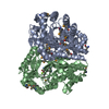
| |||||||||
|---|---|---|---|---|---|---|---|---|---|---|
| 1 |
| |||||||||
| Unit cell |
| |||||||||
| Components on special symmetry positions |
|
-Components
| #1: Protein | Mass: 45989.898 Da / Num. of mol.: 2 Source method: isolated from a genetically manipulated source Source: (gene. exp.) Citrobacter amalonaticus (bacteria) / Gene: MAL / Plasmid: PMAL / Production host: #2: Chemical |   Mass: 24.305 Da / Num. of mol.: 2 / Source method: obtained synthetically / Formula: Mg Mass: 24.305 Da / Num. of mol.: 2 / Source method: obtained synthetically / Formula: Mg#3: Chemical |   Type: L-peptide linking / Mass: 147.129 Da / Num. of mol.: 2 / Source method: obtained synthetically / Formula: C5H9NO4 Type: L-peptide linking / Mass: 147.129 Da / Num. of mol.: 2 / Source method: obtained synthetically / Formula: C5H9NO4#4: Water | ChemComp-HOH / |  Mass: 18.015 Da / Num. of mol.: 629 / Source method: isolated from a natural source / Formula: H2O Mass: 18.015 Da / Num. of mol.: 629 / Source method: isolated from a natural source / Formula: H2OHas protein modification | Y | |
|---|
-Experimental details
-Experiment
| Experiment | Method: X-RAY DIFFRACTION |
|---|
- Sample preparation
| Crystal | Density Matthews: 2.79 Å3/Da / Density % sol: 55.85 % | ||||||||||||||||||||||||
|---|---|---|---|---|---|---|---|---|---|---|---|---|---|---|---|---|---|---|---|---|---|---|---|---|---|
| Crystal grow | Temperature: 290 K / Method: vapor diffusion, hanging drop / pH: 8 Details: potassium phosphate, ammonium sulphate, pH 8.0, VAPOR DIFFUSION, HANGING DROP, temperature 290K | ||||||||||||||||||||||||
| Crystal grow | *PLUS Details: Levy, C.W., (2001) Acta Crystallogr., D57, 1922. | ||||||||||||||||||||||||
| Components of the solutions | *PLUS
|
-Data collection
| Diffraction | Mean temperature: 100 K |
|---|---|
| Diffraction source | Source: ROTATING ANODE / Type: RIGAKU RU200 / Wavelength: 1.54179 Å |
| Detector | Type: MARRESEARCH / Detector: IMAGE PLATE / Date: Jun 20, 2001 / Details: mirrors |
| Radiation | Protocol: SINGLE WAVELENGTH / Monochromatic (M) / Laue (L): M / Scattering type: x-ray |
| Radiation wavelength | Wavelength: 1.54179 Å / Relative weight: 1 |
| Reflection | Resolution: 2.1→18 Å / Num. all: 60480 / Num. obs: 60480 / Redundancy: 3 % / Rmerge(I) obs: 0.056 / Net I/σ(I): 18.2 |
| Reflection shell | Resolution: 2.1→2.15 Å / Rmerge(I) obs: 0.218 / Mean I/σ(I) obs: 5.1 / % possible all: 91.6 |
| Reflection | *PLUS Lowest resolution: 18 Å / % possible obs: 93.4 % |
| Reflection shell | *PLUS % possible obs: 91.6 % |
- Processing
| Software | Name: REFMAC / Classification: refinement | ||||||||||||||||||||
|---|---|---|---|---|---|---|---|---|---|---|---|---|---|---|---|---|---|---|---|---|---|
| Refinement | Resolution: 2.1→18 Å / SU B: 5.805 / SU ML: 0.159 / Cross valid method: THROUGHOUT / ESU R: 0.201 / ESU R Free: 0.176
| ||||||||||||||||||||
| Displacement parameters | Biso mean: 25.34 Å2
| ||||||||||||||||||||
| Refinement step | Cycle: LAST / Resolution: 2.1→18 Å
| ||||||||||||||||||||
| Refine LS restraints |
| ||||||||||||||||||||
| LS refinement shell | Highest resolution: 2.1 Å / Rfactor Rfree: 0.254 / Rfactor Rwork: 0.184 / Total num. of bins used: 20 | ||||||||||||||||||||
| Refinement | *PLUS Highest resolution: 2.1 Å / % reflection Rfree: 5.1 % / Rfactor obs: 0.167 / Rfactor Rfree: 0.227 | ||||||||||||||||||||
| Solvent computation | *PLUS | ||||||||||||||||||||
| Displacement parameters | *PLUS | ||||||||||||||||||||
| Refine LS restraints | *PLUS
|
