 Movie
Movie Controller

[English] 日本語
 Yorodumi
- PDB-6bon: Crystal structure of 2-methylcitrate synthase from Aspergillus fu... -
+ Open data

- Basic information
| Entry | Database: PDB / ID: 6bon | ||||||
|---|---|---|---|---|---|---|---|
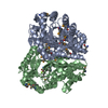
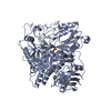
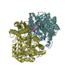
| Title | Crystal structure of 2-methylcitrate synthase from Aspergillus fumigatus with oxaloacetate and coenzyme-A. | ||||||
 Components Components | 2-methylcitrate synthase, mitochondrial | ||||||
 Keywords Keywords | TRANSFERASE / mcsA / 2-methylcitrate synthase / citrate synthase | ||||||
| Function / homology |  Function and homology information Function and homology information2-methylcitrate synthase / 2-methylcitrate synthase activity / : / citrate synthase (unknown stereospecificity) / citrate synthase activity / tricarboxylic acid cycle / carbohydrate metabolic process / mitochondrial matrix / identical protein binding Similarity search - Function | ||||||
| Biological species |  | ||||||
| Method |  X-RAY DIFFRACTION / SYNCHROTRON / MOLECULAR REPLACEMENT / Resolution: 2.35 Å X-RAY DIFFRACTION / SYNCHROTRON / MOLECULAR REPLACEMENT / Resolution: 2.35 Å | ||||||
 Authors Authors | Schlachter, C. / Chruszcz, M. | ||||||
 Citation Citation | Journal: Biol.Chem. / Year: 2019 Title: Comparative studies of Aspergillus fumigatus 2-methylcitrate synthase and human citrate synthase. Authors: Schlachter, C.R. / Klapper, V. / Radford, T. / Chruszcz, M. | ||||||
| History |
|
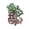
- Structure visualization
| Structure viewer | Molecule: MolmilJmol/JSmol |
|---|
- Downloads & links
-Download
| PDBx/mmCIF format | 6bon.cif.gz | 340.9 KB | Display | PDBx/mmCIF format |
|---|---|---|---|---|
| PDB format | pdb6bon.ent.gz | 277.3 KB | Display | PDB format |
| PDBx/mmJSON format | 6bon.json.gz | Tree view | PDBx/mmJSON format | |
| Others |  Other downloads Other downloads |
-Validation report
| Arichive directory | https://data.pdbj.org/pub/pdb/validation_reports/bo/6bonftp://data.pdbj.org/pub/pdb/validation_reports/bo/6bon | HTTPS FTP |
|---|
-Related structure data
| Related structure data | 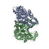 5uqoSC 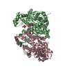 5uqqC 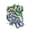 5uqrC 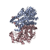 5uqsC 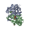 5uquC 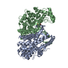 5uzpC 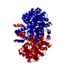 5uzqC  5uzrC  6bolC 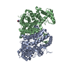 6bomC  6booC 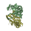 6bopC S: Starting model for refinement C: citing same article ( |
|---|---|
| Similar structure data |


-Links
 PDBj
PDBj


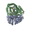
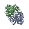
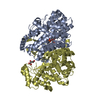
- Assembly
| Deposited unit | 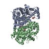
| ||||||||||||||||||
|---|---|---|---|---|---|---|---|---|---|---|---|---|---|---|---|---|---|---|---|
| 1 |
| ||||||||||||||||||
| Unit cell |
| ||||||||||||||||||
| Noncrystallographic symmetry (NCS) | NCS domain:
NCS domain segments: Component-ID: _ / Ens-ID: 1 / Beg auth comp-ID: ALA / Beg label comp-ID: ALA / End auth comp-ID: LYS / End label comp-ID: LYS / Refine code: _ / Auth seq-ID: 31 - 464 / Label seq-ID: 7 - 440
|
-Components
| #1: Protein | Mass: 48557.426 Da / Num. of mol.: 2 Source method: isolated from a genetically manipulated source Source: (gene. exp.) Strain: CEA10 / CBS 144.89 / FGSC A1163 / Gene: mcsA, AFUB_094700 / Production host:  References: UniProt: B0YD89, 2-methylcitrate synthase, citrate synthase (unknown stereospecificity) #2: Chemical |   Mass: 131.064 Da / Num. of mol.: 3 / Source method: obtained synthetically / Formula: C4H3O5 Mass: 131.064 Da / Num. of mol.: 3 / Source method: obtained synthetically / Formula: C4H3O5#3: Chemical | ChemComp-COA / |   Mass: 767.534 Da / Num. of mol.: 1 / Source method: obtained synthetically / Formula: C21H36N7O16P3S Mass: 767.534 Da / Num. of mol.: 1 / Source method: obtained synthetically / Formula: C21H36N7O16P3S#4: Water | ChemComp-HOH / |  Mass: 18.015 Da / Num. of mol.: 145 / Source method: isolated from a natural source / Formula: H2O Mass: 18.015 Da / Num. of mol.: 145 / Source method: isolated from a natural source / Formula: H2O |
|---|
-Experimental details
-Experiment
| Experiment | Method: X-RAY DIFFRACTION / Number of used crystals: 1 |
|---|
- Sample preparation
| Crystal | Density Matthews: 2.06 Å3/Da / Density % sol: 40.41 % |
|---|---|
| Crystal grow | Temperature: 298 K / Method: vapor diffusion, sitting drop / pH: 6.5 Details: 0.1 M Bis-Tris pH 6.5, 28% PEGMME2000 1.0 mM oxaloacetate 1.0 mM coenzyme-A 1.0 mM Guanosine monophosphate |
-Data collection
| Diffraction | Mean temperature: 100 K |
|---|---|
| Diffraction source | Source: SYNCHROTRON / Site: APS  / Beamline: 22-ID / Wavelength: 1 Å / Beamline: 22-ID / Wavelength: 1 Å |
| Detector | Type: RAYONIX MX300-HS / Detector: CCD / Date: Oct 16, 2016 |
| Radiation | Protocol: SINGLE WAVELENGTH / Monochromatic (M) / Laue (L): M / Scattering type: x-ray |
| Radiation wavelength | Wavelength: 1 Å / Relative weight: 1 |
| Reflection | Resolution: 2.35→50 Å / Num. obs: 32572 / % possible obs: 96.1 % / Observed criterion σ(I): -3 / Redundancy: 4.7 % / Rmerge(I) obs: 0.077 / Rpim(I) all: 0.052 / Rrim(I) all: 0.119 / Rsym value: 0.077 / Net I/σ(I): 14.5 |
| Reflection shell | Resolution: 2.35→2.39 Å / Redundancy: 4.7 % / Rmerge(I) obs: 0.524 / Mean I/σ(I) obs: 2.35 / Num. unique obs: 1649 / Rpim(I) all: 0.308 / Rrim(I) all: 0.678 / Rsym value: 0.524 / % possible all: 99.1 |
- Processing
| Software |
| ||||||||||||||||||||||||||||||||||||||||||||||||||||||||||||||||||||||||||||||||||||||||||||||||||||||||||||||||||||||||||||||||||||||||||||||||||||||||||||||||||||||||||||||||||||||
|---|---|---|---|---|---|---|---|---|---|---|---|---|---|---|---|---|---|---|---|---|---|---|---|---|---|---|---|---|---|---|---|---|---|---|---|---|---|---|---|---|---|---|---|---|---|---|---|---|---|---|---|---|---|---|---|---|---|---|---|---|---|---|---|---|---|---|---|---|---|---|---|---|---|---|---|---|---|---|---|---|---|---|---|---|---|---|---|---|---|---|---|---|---|---|---|---|---|---|---|---|---|---|---|---|---|---|---|---|---|---|---|---|---|---|---|---|---|---|---|---|---|---|---|---|---|---|---|---|---|---|---|---|---|---|---|---|---|---|---|---|---|---|---|---|---|---|---|---|---|---|---|---|---|---|---|---|---|---|---|---|---|---|---|---|---|---|---|---|---|---|---|---|---|---|---|---|---|---|---|---|---|---|---|
| Refinement | Method to determine structure: MOLECULAR REPLACEMENT Starting model: 5UQO Resolution: 2.35→50 Å / Cor.coef. Fo:Fc: 0.943 / Cor.coef. Fo:Fc free: 0.91 / Cross valid method: THROUGHOUT / ESU R: 0.748 / ESU R Free: 0.295 / Details: HYDROGENS HAVE BEEN ADDED IN THE RIDING POSITIONS
| ||||||||||||||||||||||||||||||||||||||||||||||||||||||||||||||||||||||||||||||||||||||||||||||||||||||||||||||||||||||||||||||||||||||||||||||||||||||||||||||||||||||||||||||||||||||
| Solvent computation | Ion probe radii: 0.8 Å / Shrinkage radii: 0.8 Å / VDW probe radii: 1.2 Å | ||||||||||||||||||||||||||||||||||||||||||||||||||||||||||||||||||||||||||||||||||||||||||||||||||||||||||||||||||||||||||||||||||||||||||||||||||||||||||||||||||||||||||||||||||||||
| Displacement parameters | Biso mean: 37.116 Å2
| ||||||||||||||||||||||||||||||||||||||||||||||||||||||||||||||||||||||||||||||||||||||||||||||||||||||||||||||||||||||||||||||||||||||||||||||||||||||||||||||||||||||||||||||||||||||
| Refinement step | Cycle: 1 / Resolution: 2.35→50 Å
| ||||||||||||||||||||||||||||||||||||||||||||||||||||||||||||||||||||||||||||||||||||||||||||||||||||||||||||||||||||||||||||||||||||||||||||||||||||||||||||||||||||||||||||||||||||||
| Refine LS restraints |
|